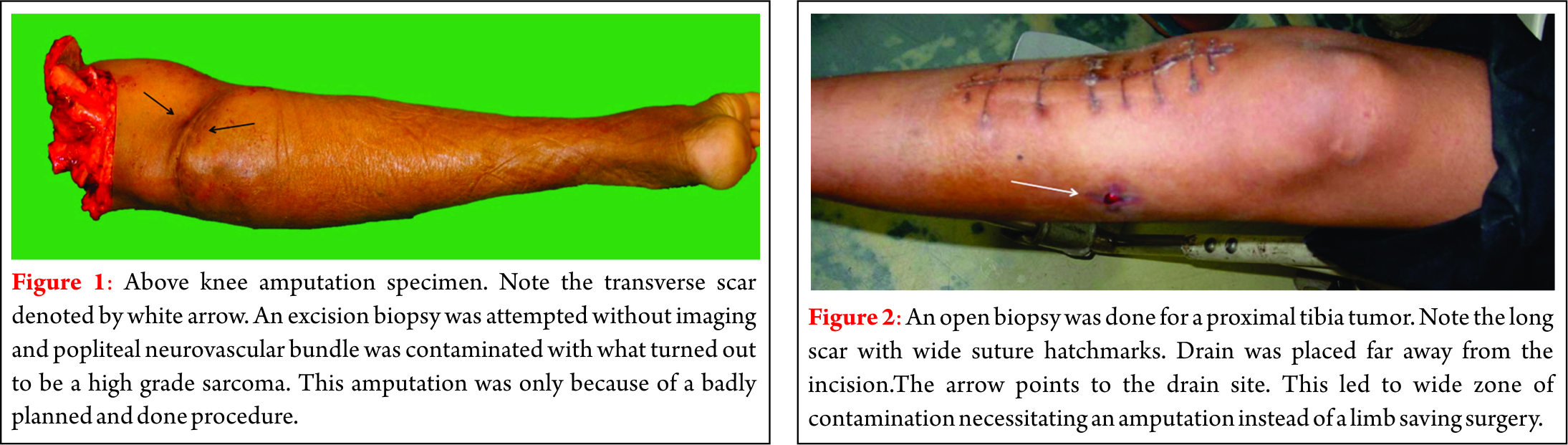
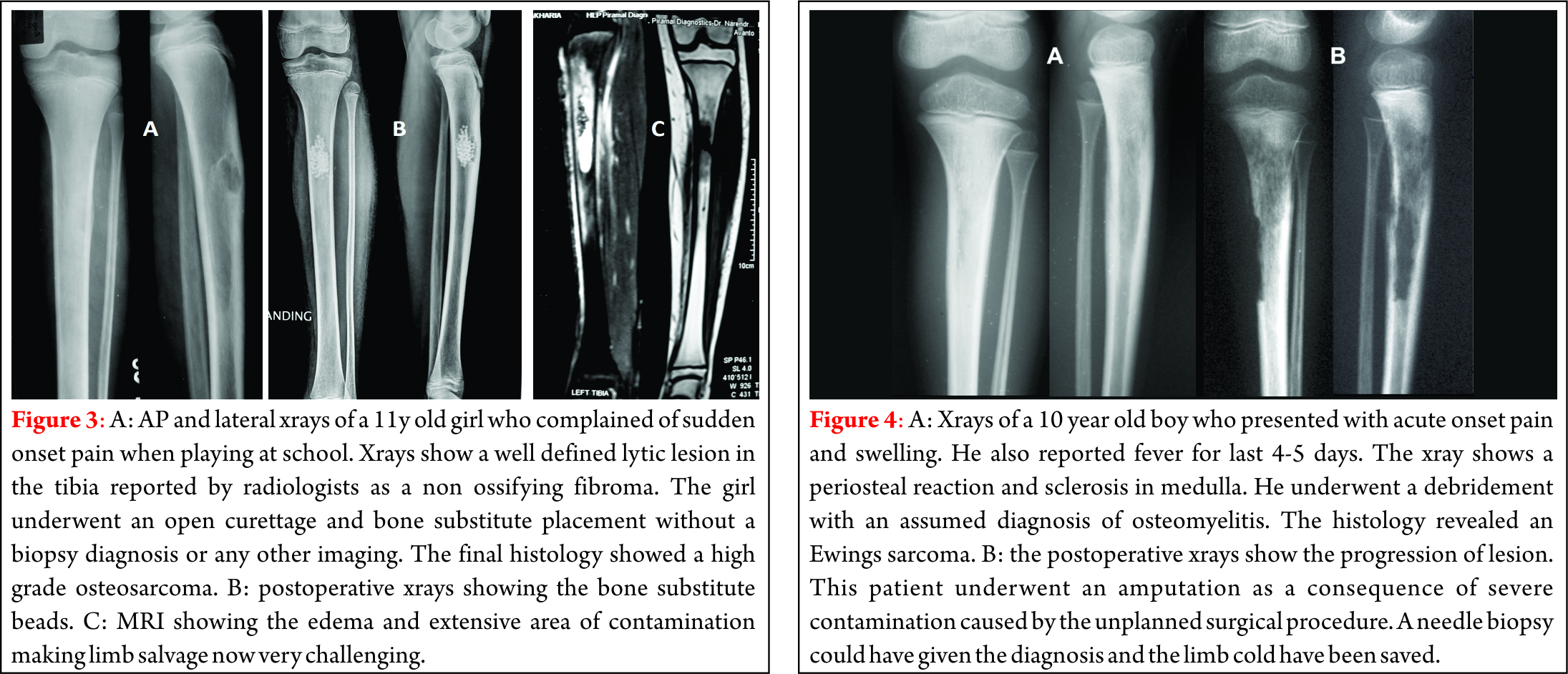
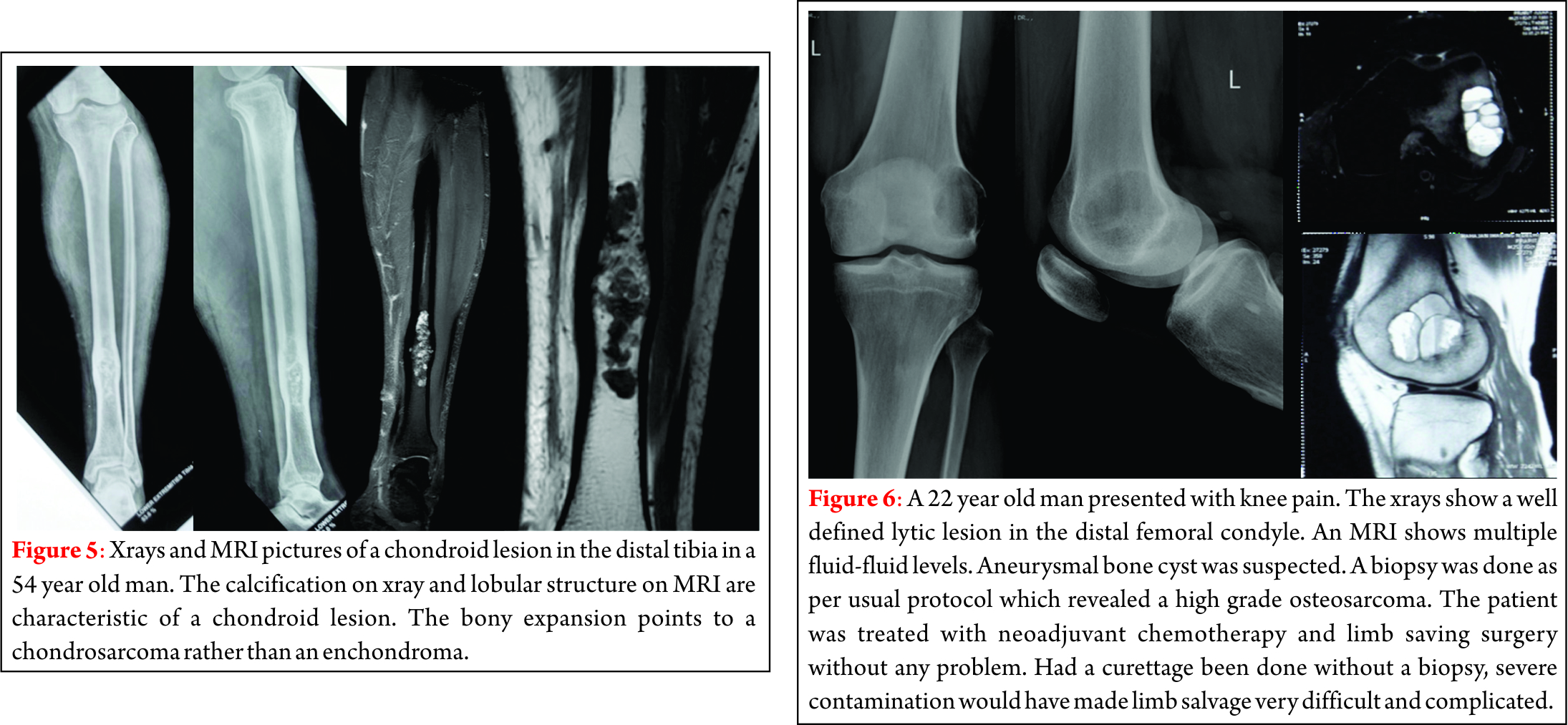
Lumbar Prolapsed Intervertebral Disc – A Treatment Algorithm

Vol 1 | Issue 1 |  July – Dec 2016 | Page 29-35 | Akshay Gadiya, Mandar Borde, Priyank Patel, Shekhar Bhojraj, Premik Nagad, Tanay Prabhoo.


Authors: Akshay Gadiya [1], Mandar Borde [1], Priyank Patel [1], Shekhar Bhojraj [1], Premik Nagad [1], Tanay Prabhoo [1].

[1] The Spine Clinic, Lilavati Hospital and Research Centre, Bandra West, Mumbai, India..

Address of Correspondence
Dr. Akshay Gadiya,
The Spine Clinic, Lilavati Hospital and Research Centre,
Bandra West, Mumbai.
E-mail: akshaygadia@gmail.com


Abstract

Lumbar prolapsed intervertebral disc (PID) or herniations is the most common cause of spine related disability in working-age individuals. Symptomatic prolapsed disc presents as lumbar radiculopathy due to both mechanical compression as well as chemical irritation of nerve root. It is common problem encountered in both surgical and non-surgical practice. There is variety of non- surgical as well as surgical treatment available for treating this common ailment. Though various protocols are available to promote improved outcome and cost effectiveness and reduce unnecessary interventions the decision between surgical and non-surgical management of this entity can be challenging to both patient and treating physician. The aim of this article is to discuss the etiology and pathophysiology of lumbar PID and provide a practical and evidence based algorithm for its treatment.
Keywords: Lumbar prolapsed intervertebral disc, management algorithm


Background
Lumbar PID can be extremely painful and cause significant morbidity and loss of function [1,2]. It can lead to substantial radicular symptoms, which if persistent can lead to surgical intervention. Intervertebral disc in lumbar spine are complex structures that are subjected to significant axial loading along with shearing forces.[1] Because of these biomechanical demands along with the inability to remodel owing to avascular nature, herniations of lumbar intervertebral discs are common. The armamentarium of treatment for this pathology extends from simple conservative treatment of complete bed rest to highly complex percutaneous endoscopic microdiscectomy[3]. Indirect cost associated with this condition may include work absenteeism, short and long term disability and reduced work capacity secondary to pain and/or weakness. Fortunately 90% of cases of sciatica from herniated lumbar disc resolve in about 12 weeks[4]. In the absence of progressive neurological deficits or cauda equina syndrome, non-surgical treatments are implemented for 6 weeks with good results[2]. Conservative treatment is recommended to reduce pain and improve function in this time period while the body hopefully will resorb the disc material. Surgical treatment is indicated in scenario of worsening neurological deficit, cauda equina syndrome and failure of conservative treatment.

Anatomy of Intervertebral Disc:
Intervertebral disc is composed of cartilaginous end plates, the annulus fibrosus (AF), and the nucleus pulposus (NP)[5]. The end plates are intermittent structures between the subchondral bone of vertebral body and the AF of intervertebral disc. The end plates are made up of 1-mm thick layer of hyaline cartilage, which is comprised of 50% water, chondrocytes, proteoglycans (PGs), and type II collagen [6]. An extensive capillary network exists in end plate that extends one to two millimeters in AF. This vascular network is responsible for providing nutrition to the otherwise avascular intervertebral disc.
AF forms the outer ring of intervertebral disc forming 15-25 lamellar rings and is composed primarily of fibroblast like cells and type 1 collagen fibers*. It is commonly divided into outer and inner AF [7]. The outer layer is highly organized and almost exclusively made of type I collagen, resulting in high tensile strength. Comparatively the inner layer is a transitional zone between the AF and NP and has both type I and type II collagen, as well as multiple different proteoglycans[8]. NP is the central jelly like substance composed primarily of chondrocyte like cells that are responsible for secreting type II collagen as well as numerous PGs. Aggrecan is the most common PG in NP and is responsible for its substantial hydrophilic nature[7]. NP is responsible for the ability of IVD to withstand the substantial axial loads. Recently notochordal cells have been identified in the NP that is responsible for preventing the apoptosis of chondrocyte like cells [9].

Types of lumbar disc herniation:
While often times the terms disc herniation, disc protrusion, and disc bulge are used interchangeably in the literature, according to the combined task forces of the North American Spine Society, the American Society of Spine Radiology, and the American Society of Neuroradiology, these pathologies are not the same; they define a disc herniation as “localized or focal displacement of disc material beyond the limits of the intervertebral disc space”[10]. This differentiation is critical, as diffuse enlargement of annulus cannot be labeled as true herniation as it is a variant of disc degeneration. A true herniated disc is a focal pathology that affects less than 25% of the intervertebral disc[10]. Herniated discs can be categorized as protrusions, extrusions, or sequestrations (Fig. 1). Protrusions are wide-based herniations in which the diameter at the base of the herniation is wider than the diameter of the herniation in the canal. Extrusions have a narrow base, with a large herniation in the canal, and sequestrations are herniations in which there is no continuity between the herniation and the remaining intervertebral disc[10].

Figure 1: Types of lumbar disc herniation. Protrusion (A), Extrusion (B) and Sequestration (C).

Figure 1: Types of lumbar disc herniation. Protrusion (A), Extrusion (B) and Sequestration (C).

Epidemiology of Lumbar PID:
Extensive research has been performed into epidemiology of lumbar disc herniations till date and many possible risk factors have been identified. Cummins et al.[11]reported that the average age of patients with a herniated disc was 41 years, and the diagnosis was slightly more common in males than females (57% versus 43%, respectively).
Obesity is a major comorbidity associated with lumbar disc herniations. An elevated body mass index (BMI) thought to increase the axial load on lumbar spine resulting in herniated intervertebral disc[12]. Bostman reported 27% of the patients undergoing surgery for a lumbar disc herniation were obese, whereas the population prevalence of obesity in Finland at that time was only 16%. A recent meta-analysis by Shiri et al. found that overweight patients (BMI: 25–30) and obese patients (BMI > 30) had a statistically significant increase risk of being diagnosed with lumbar radiculopathy than patients with a BMI < 25[13].
Other medical comorbidities such as diabetes, hyperlipidemia, and smoking have also been reported as possible risk factors for lumbar disc herniations. Sakellaridis compared a case series of 102 patients requiring surgical intervention for a lumbar disc herniation to 98 patients undergoing elective surgery for another pathology and found a statically significant increase in the rate of diabetes in patients undergoing a lumbar microdiscectomy (32% versus 19%, p = 0.001). Also Mobbs et al.[14] reported that the need for revision surgery for diabetic patients was 7 times higher than non- diabetic patients. In a meta-analysis of 49 articles, Jordan et al. identified smoking as an independent risk factor for lumbar disc herniations[15]. Although the mechanism by which this comorbidities increase the rate of lumbar disc herniations is yet to be proven it is speculated that there is either decrease in microcirculation or increase in cytokine expression.
Occupational risk factors have been demonstrated as a major risk factor for lumbar disc herniations as this pathology is more common in working age individuals. Cummins et al.[11] reported that 20% of the patients with lumbar disc herniation had a workers’ compensation claim, compared to only 8% of patients with spinal stenosis and 7% of the patients with a degenerative spondylolisthesis. In a multi-center, case–control study, Seidler et al.[16] found a dose–response relationship between the total work-related lumbar load and the incidence of lumbar disc herniations. Multiple biomechanical studies have demonstrated that the combination of an axial load and twisting mechanism or an axial load and a flexion mechanism can lead to a herniated disc[17].
Finally, there is a clear genetic link established for lumbar disc herniations and lumbar disc degenerations[18]. Zhang et al.[19], in a case control study of 4000 patients, reported that a family history of a lumbar disc herniation was the most important risk factor in predicting patients who would develop a lumbar disc herniation (odds ratio 3.6). Although this etiology is multifactorial, the collagen IX tryptophan allele (Trp2) has been linked to an increased severity of disc degeneration in patients less than 40 years of age with a lumbar disc herniation[20].

Pathophysiology:
While a herniated disc is traditionally thought of as a herniation of the NP through the AF, histologic examination of surgical specimens has shown that a pure herniation of NP is rare. The annulus makes up a portion of the herniation in two-thirds of the cases, and approximately 20% of all herniations include a portion of the cartilaginous endplate[7]. Lumbar disc herniations are most commonly posterolateral herniations that affect the traversing nerve root, and pain may either be from mechanical compression or chemical irritation of the nerve root. Mechanical compression can deform and stretch the nerve, as well as compress the microcirculation leading to ischemia and radicular symptoms; additionally, the herniation stimulates a substantial inflammatory cascade that is critical in the resorption of the disc herniation, but it can also lead to chemical irritation of the nerve root and radicular symptoms[21]. While the increase of number of cytokines have been described after lumbar disc herniations, the role of only few have cytokines on disc resorption and radicular symptoms have been established[22]. The concentration of FGF is elevated in surgical specimens of human lumbar disc herniations, and this cytokine potently attracts macrophages to the injury site[23]. While the inflammatory cascade is beneficial because it leads to resorption of the herniated discs, it also is partly responsible for the symptoms from a herniated disc. While there is little innervation to uninjured intervertebral discs, after being exposed to the inflammatory cascade, up to 80% of disc herniations have nerves present after being surgically removed[7]. Additionally, there is a significant increase in the concentration of TNF alpha in lumbar disc herniations compared to intact intervertebral discs[24], and TNF alpha has been repeatedly demonstrated to stimulate radiculitis[25].

Natural History of lumbar PID:
The decision to treat any condition depends upon an understanding of the natural history of the disease process. The natural history of a lumbar herniation of the nucleus pulposus (HNP) is not fully known and clear indications for operative intervention cannot be established from the literature. Controversy still exists regarding indications for operating on large, extruded discs. A large extruded disc has been a relative indication for operative treatment in the past[26]. However, several papers have demonstrated that these discs have the greatest tendency to decrease in size with conservative management[4,27]. The Spine Patient Outcomes Research Trial (SPORT)[28] recruited 501 surgical candidates and randomized them into discectomy versus conservative treatment. At 2-year follow-up, both groups had made substantial improvement. There was a tendency towards better outcomes in the operative group but this was not statistically significant. A large crossover of patients between the groups hampered the study. In a quest to identify the natural history of massive herniated discs, Benson et al.[29] retrospectively studied 34 patients over a period of 7 years. They concluded that where clinical progress is evident, 83 % cases of massive disc herniation will have sustained improvement and it is safe to adopt ‘wait and watch’ policy in cases of massive disc herniation if there is early sign of clinical recovery.

Non-operative management:
As described in a previous section, more than 80 % cases with lumbar disc herniations have favourable outcome when left untreated. This fact underlines the role of non-operative treatment in the management of lumbar disc herniations. Conservative treatment is recommended to reduce pain and improve function in this time period while the body hopefully will resorb the disc material. Several conservative options exist, but the data is unclear as to which are truly efficacious. Numerous medications, including acetaminophen, non- steroidal anti-inflammatory drugs (NSAIDs), muscle relaxants, steroids, narcotics, neuroleptics, and anti-depressants, are used to treat back pain and radicular symptoms that result from lumbar disc herniation. NSIADs are often utilized as a first-line treatment, but there is limited data supporting their benefit[30]. Oral corticosteroids are also commonly prescribed for acute disc herniations and lower back pain, but data regarding their efficacy is limited[31]. Membrane-stabilizing agents such as gabapentin and pregabalin show modest benefit[32].
Spinal Manipulative therapy (SMT) is another mode of non-operative treatment used by Osteopathic physicians, chiropractors and physical therapists[33]. Quality studies on the efficacy of SMT in the treatment of symptomatic lumbar herniated discs are lacking[34]. Recent review is unable to make strong recommendations for or against its use in low-back and radicular pain due to poor quality evidence[35].
The use of epidural steroids for treatment of sciatica was first documented in 1952. Since then the use of epidural steroid injections for treatment of symptomatic lumbar disc herniations has increased exponentially and has become a commonplace in treatment of symptomatic lumbar disc herniations. The use of fluoroscopy has allowed for more accurate and safe placement of injectate in epidural space. Saal [37] first demonstrated the rational explanation for use of corticosteroids in 1990. He demonstrated the elevated levels of Phospholipase A2 at the interference between herniated nucleus pulposus and nerve root. There are 3 techniques for injection in to epidural space namely caudal approach, interlaminar approach and the transforaminal approach. The discussion on details of techniques is beyond the scope of this article. However, transforaminal approach is used most frequently. Schaufele et al.[37] compared transforaminal to interlaminar epidural injections for lumbar disc herniation and found that the transforaminal injection group had greater improvement than the interlaminar group.
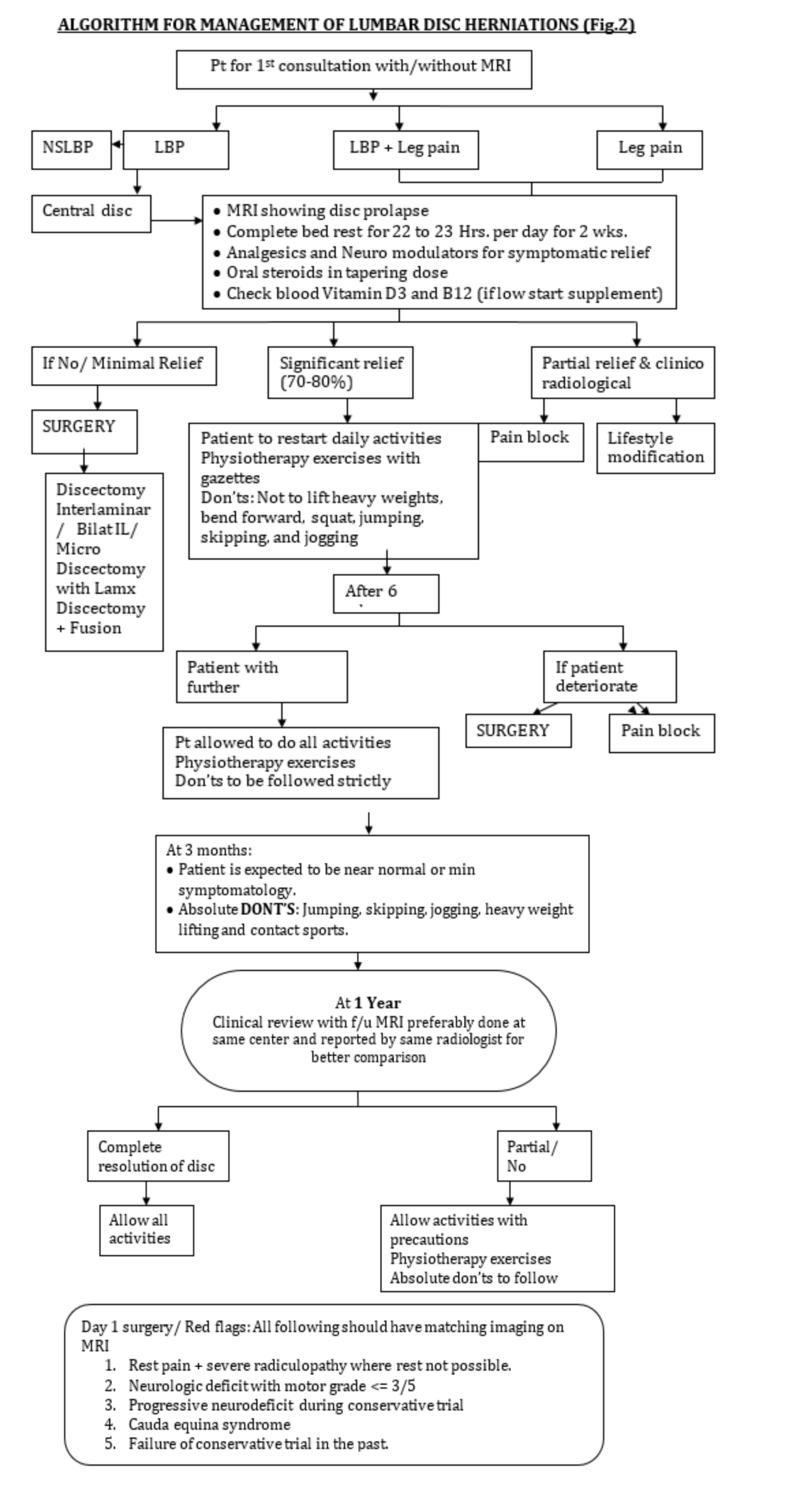
In our practice, we prescribe complete bed rest for a period of 2 weeks. We believe off loading the spine help in early resolution of acute symptoms. We have also experienced that a short course of intravenous methyl prednisolone at 1g/day for 3 days help to reduce chemical radiculitis occurring because of herniated disc. After 2 weeks patient is asked to mobilize out of bed and undergo a systematic physiotherapy program for another 3 weeks. Patient is asked to assess the claudication and to keep a watch for ‘red flag’ signs. In case any of latter things occur or the claudication distance is progressively decreasing, the patient follows up immediately and is evaluated for surgical management (Fig.2). Patients who are not significantly better symptomatically at the end of 2 weeks after complete bed rest are also the candidates for surgical management.

8-pid-fig-2

Operative treatment:
The absolute indications for surgical treatment in patients with lumbar disc herniations are worsening neurological deficit and cauda equina syndrome. Latter is a surgical emergency and is characterized by perianal sensory deficit, bowel and bladder incontinence and either a new or progressive deficit. More often the herniations are central and presents more frequently in men in the fifth decade of life. It is commonly found in L4-L5 disc [38]. Relative indications for surgical treatment vary and are surgeon as well as patient dependent. There are certain prerequisites that we follow before deciding on surgical treatment. Patient should have demonstrable pathology on radiology and correlative physical examination in displaying motor and sensory symptomatology in addition to failure of non-operative treatment. Operative treatment for lumbar disc herniations include endoscopic microdiscectomy, micro lumbar discectomy, interlaminar discectomy with or without foraminotomy, conventional open laminectomy and discectomy with or without instrumented fusion and disc replacement[39]. Whatever surgical option being chosen, the aim of surgery should be thorough decompression of nerve roots. It has been always the matter of debate regarding amount of disc to be removed during discectomy. Spengler[40], in 1990 in a case control study concluded that results of radical discectomy were comparable to limited discectomy. Conventional open laminectomy and discectomy is preferred in patients with co existent lumbar canal stenosis. Another important question to address is to do fusion or not do fusion along with decompression as the there are advocates for both the lines of treatment in literature[41]. Proponents of fusion describe discectomy as the destabilizing procedure and thus fusion is required to stabilize the spine[42]. However, the other school of thought believes in just adequate lumbar decompression. We belong to the second group and do not believe in prophylactic fusion. Frymoyer et al.[43] in 1978 gave the guidelines for fusion in lumbar disc herniation surgeries. Fusion was indicated in patients with acute disc herniations and protracted significant component of back pain, symptomatic and radiologically demonstrable segmental instability and presence of neural arch defects along with disc disease. Pedicle screw fixation along with intertransverse posterolateral (PLF) fusion is the modality of fusion preferred in our unit whenever we fuse the cases of lumbar disc herniations. Interbody fusion is performed only in cases having lumbar disc herniations along with reducible lytic listhesis at the same level. In a recently published study by Glassmann et al.[44], transforaminal lumbar interbody fusion (TLIF) fared better than PLF in patients of spondylolisthesis. While in other pathologies TLIF and PLF had same functional outcome. Advantage of PLF over TLIF is less surgical time and less blood loss..


Conclusion

To conclude, lumbar disc herniations are major cause of lower back-related disability in working-age group. Fortunately, around 80 % of patients do well with non-operative treatment while surgery is reserved for a small and specific fraction of patients. There is a wide range of modalities in non-operative management of lumbar disc herniations inspite of lack of evidence for any specific modality better than other. In cases of clinico radiological mismatch epidural steroids is preferred modality of treatment. Whenever an operative treatment is opted we don’t believe in prophylactic fusion. Instability should be given a chance. Fusion is performed only in limited and specific patients. Pedicle screws fixation along with posterolateral fusion (PLF) is a preferred modality of treatment.


References

1. Shroeder G, Guyre C, Vaccaro A. The epidemiology and pathophysiology of lumbar disc herniations. Semin. Spine Surgery. 2016;28 : 2-7.
2. Simon J, Conliffe T, Kitei P. Non operative managemet: An evidence based approach. Semin. Spine Surgery.2016;28: 8-13.
3. Schoenfeld AJ, Weiner BK. Treatment of lumbar disc herniation: Evidence-based practice. Int J Gen Med. 2010 Jul 21;3:209-14
4. Saal JA, Saal JS. Nonoperative treatment of herniated lumbar intervertebral disc with radiculopathy. An outcome study. Spine. 1989;14(4):431–437.
5. Colombier P, Clouet J, Hamel O, Lescaudron L, Guicheux J. The lumbar intervertebral disc: from embryonic development to degeneration. Joint Bone Spine. 2014;81(2):125–129.
6. Roberts S, Menage J, Urban JP. Biochemical And Structural Properties Of The Cartilage End-Plate And Its Relation To The Intervertebral Disc. Spine. 1989;14(2):166–174.
7. Roberts S, Evans H, Trivedi J, Menage J. Histology and pathology of the human intervertebral disc. J Bone Joint Surg Am. 2006;88(suppl 2):10–14.
8. Eyre DR, Muir H. Types I and II collagens in intervertebral disc. Interchanging radial distributions in annulus fibrosus. Bio- chem J. 1976;157(1):267–270.
9. Erwin WM, Islam D, Inman RD, Fehlings MG, Tsui FW. Notochordal cells protect nucleus pulposus cells from degra- dation and apoptosis: implications for the mechanisms of intervertebral disc degeneration. Arthritis Res Ther. 2011;13(6): R215.
10. Fardon DF, Williams AL, Dohring EJ, Murtagh FR, Gabriel Rothman SL, Sze GK. Lumbar disc nomenclature: version 2.0: recommendations of the combined task forces of the North American Spine Society, the American Society of Spine Radiology and the American Society of Neuroradiology. Spine J. 2014;14(11):2525–2545.
11. Cummins J, Lurie JD, Tosteson TD, et al. Descriptive epidemi- ology and prior healthcare utilization of patients in the Spine Patient Outcomes Research Trial’s (SPORT) three observatio- nal cohorts: disc herniation, spinal stenosis, and degenerative spondylolisthesis. Spine. 2006;31(7):806–814.
12. Weiler C, Lopez-Ramos M, Mayer HM, et al. Histological analysis of surgical lumbar intervertebral disc tissue provides evidence for an association between disc degeneration and increased body mass index. BMC Res Notes. 2011;4(4):497.
13. Shiri R, Lallukka T, Karppinen J, Viikari-Juntura E. Obesity as a risk factor for sciatica: a meta-analysis. Am J Epidemiol. 2014;179(8):929–937.
14. Mobbs RJ, Newcombe RL, Chandran KN. Lumbar discectomy and the diabetic patient: incidence and outcome. J Clin Neuro- sci. 2001;8(1):10–13.
15. Jordan J, Konstantinou K, O’Dowd J. Herniated lumbar disc. BMJ Clin Evid. 2009;3(3):1118.
16. Seidler A, Bergmann A, Jager M, et al. Cumulative occupa- tional lumbar load and lumbar disc disease—results of a German multi-center case–control study (EPILIFT). BMC Mus- culoskelet Disord. 2009;10(1):48.
17. Palmer KT, Griffin M, Ntani G, et al. Professional driving and prolapsed lumbar intervertebral disc diagnosed by magnetic resonance imaging: a case–control study. Scand J Work Environ Health. 2012;38(6):577–581.
18. Battie MC, Videman T, Kaprio J, et al. The twin spine study: 
contributions to a changing view of disc degeneration. Spine J. 
2009;9(1):47–59.
19. Zhang YG, Sun Z, Zhang Z, Liu J, Guo X. Risk factors for 
lumbar intervertebral disc herniation in Chinese population: 
a case–control study. Spine. 2009;34(25):E918–E922.
20. Higashino K, Matsui Y, Yagi S, et al. The alpha2 type IX collagen tryptophan polymorphism is associated with the severity of disc degeneration in younger patients with herni- ated nucleus pulposus of the lumbar spine. Int Orthop. 2007;31 
(1):107–111.
21. Kawaguchi S, Yamashita T, Yokogushi K, Murakami T, Ohwada O, Sato N. Immunophenotypic analysis of the inflammatory infiltrates in herniated intervertebral discs. Spine. 2001;26(11):1209–1214.
22. Zhou G, Dai L, Jiang X, et al. Effects of human midkine on spontaneous resorption of herniated intervertebral discs. Int Orthop. 2010;34(1):103–108.
23. Doita M, Kanatani T, Harada T, Mizuno K. Immunohistologic study of the ruptured intervertebral disc of the lumbar spine. Spine. 1996;21(2):235–241.
24. Roberts S, Evans H, Menage J, et al. TNFalpha-stimulated gene product (TSG-6) and its binding protein, IalphaI, in the human intervertebral disc: new molecules for the disc. Eur Spine J. 2005;14(1):36–42.
25. Andrade P, Visser-Vandewalle V, Philippens M, et al. Tumor necrosis factor-alpha levels correlate with postoperative pain severity in lumbar disc hernia patients: opposite clinical effects between tumor necrosis factor receptor 1 and 2. Pain. 2011;152(11):2645–2652.
26. Postacchini F. Results of surgery compared with conservative management for lumbar disc herniations. Spine 1996; 21: 1383–7
27. Cribb GL, Jaffray DC, Cassar-Pullicino VN. Observations on the natural history of massive lumbar disc herniation. J Bone Joint Surg Br 2007; 89: 782–4.
28. Weinstein JN, Tosteson TD, Lurie JD, Tosteson AN, Hanscom B, Skinner JS et al. Surgical vs nonoperative treatment for lumbar disk herniation: the Spine Patient Outcomes Research Trial (SPORT): a randomized trial. JAMA 2006; 296: 2441–50.
29. Benson SP, Tavares SP, Robertson SC, Sharp P, Marshall RW. Ann R Coll Surg Engl 2010; 92: 147–153
30. Roelofs PD, Deyo RA, Koes BW, Scholten RJ, van Tulder MW. Non-steroidal anti-inflammatory drugs for low back pain. Cochrane Database Syst Rev. 2008(1):CD000396.
31. Eskin B, Shih RD, Fiesseler FW, et al. Prednisone for emer- gency department low back pain: a randomized controlled trial. J Emerg Med. 2014;47(1):65–70.
32. Yildirim K, Deniz O, Gureser G, et al. Gabapentin monother- apy in patients with chronic radiculopathy: the efficacy and impact on life quality. J Back Musculoskelet Rehabil. 2009;22(1): 17–20.
33. Assendelft WJ, Morton SC, Yu EI, Suttorp MJ, Shekelle PG. Spinal manipulative therapy for low back pain. A meta- analysis of effectiveness relative to other therapies. Ann Intern Med. 2003;138(11):871–881.
34. Rubinstein SM, van Middelkoop M, Assendelft WJ, de Boer MR, van Tulder MW. Spinal manipulative therapy for chronic low- back pain: an update of a Cochrane review. Spine. 2011;36(13): E825–E846.
35. Rubinstein SM, Terwee CB, Assendelft WJ, de Boer MR, van Tulder MW. Spinal manipulative therapy for acute low back pain: an update of the cochrane review. Spine. 2013;38(3):E158–E177.
36. Saal JS, Franson RC, Dobrow R, Saal JA, White AH, Goldthwaite N. High levels of inflammatory phospholipase A2 activity in lumbar disc herniations. Spine. 1990;15(7): 674–678.
37. Schaufele MK, Hatch L, Jones W. Interlaminar versus trans- foraminal epidural injections for the treatment of sympto- matic lumbar intervertebral disc herniations. Pain Physician. 2006;9(4):361–366.
38. Walker JL, Schulak D, Murtagh R. Midline disc herniations of the lumbar spine. South Med J. 1993;86:13–17.
39. Jacobs WC, van Tulder M, Arts M, Rubinstein SM, van Middelkoop M, Ostelo R, Verhagen A, Koes B, Peul WC. Surgery versus conservative management of sciatica due to a lumbar herniated disc: a systematic review. Eur Spine J. 2011 Apr;20(4):513-22.
40. Spengler M, Ouellette EA, Batti M, Zeh J. Elective discectomy for herniation of lumbar disc. Additional experience with an objective method. J Bone Joint Surg (Am) 1990; 72 (2): 230-7.
41. Rainville J, Lopez E. Comparison of radicular symp- toms caused by lumbar disc herniation and lumbar spinal stenosis in the elderly. Spine (Phila Pa 1976) 2013;38:1282-7.
42. Goldner JL: The role of spine fusion: question 6. Spine 6:293, 1981.
43. Frymoyer JW, Hanley E, Howe J, Kuhlmann D, Matteri R. Disc excision and spine fusion in the management of lumbar disc disease. A minimum ten-year followup. Spine (Phila Pa 1976). 1978 Mar;3(1):1-6.
44. Glassman SD, Carreon LY, Ghogawala Z, Foley KT, McGirt MJ, Asher AL. Benefit of Transforaminal Lumbar Interbody Fusion vs Posterolateral Spinal Fusion in Lumbar Spine Disorders: A Propensity-Matched Analysis From the National Neurosurgical Quality and Outcomes Database Registry. Neurosurgery. 2016 Sep;79(3):397-405.


How to Cite this article: Gadiya A, Borde M, Patel P, Bhojraj S, Nagad P, Prabhoo T. Lumbar Prolapsed intervertebral Disc – A treatment algorithm. Journal of Clinical Orthopaedics July – Dec 2016; 1(1):29-35..

(Abstract    Full Text HTML)      (Download PDF)


Biopsy for Musuloskeletal Tumors – An Orthopaedic Surgeons Guide

Vol 1 | Issue 1 |  July – Dec 2016 | Page 21-28 | Manish Agarwal.


Authors: Manish Agarwal [1].

[1] Department of Surgical Oncology P.D Hinduja Hospital & MRC Veer Savarkar Marg, Mahim, Mumbai 400016.

Address of Correspondence
Dr Manish Agarwal
Ullas, 1st floor 17 Laburnum Road, Gamdevi, Mumbai 400007.
Email: mgagarwal@gmail.com


Abstract

Introduction: Biopsy has a central role in diagnosis of musculoskeletal tumors. Inappropriately done biopsy is one of the most important causes of compromised limb salvage or worse loss of limb or life. It also has serious medicolegal implications. Problems can be avoided if principles and specially the technique of biopsy is understood clearly. This article focusses on basic principles and also provides easy guidelines for a biopsy.
Key Words: Musculoskeletal tumors, needle biopsy, open biopsy, guidelines.


Introduction

A biopsy is perhaps the most important step in the workup of bone tumors. A histological diagnosis establishes the identity of the neoplasm before any treatment is offered. In a way it is like the final conclusive identification of the “criminal” where clinical and imaging clues help us narrow down the possibilities. We are living in a modern world where sarcomas have a cure rate of 60-70% and almost 90% of these are suitable for limb salvage. Most patients are first seen by a general orthopedic surgeon who then has the responsibility of establishing the diagnosis and guiding the patient to a correct specialized center for treatment. Inappropriately done biopsy is today the commonest cause of compromised limb salvage or worse, an amputation. (Fig. 1 & 2) This also opens the door to medicolegal dispute. Mankin [1] reported a 15.9% complication rate with 3% unnecessary amputations in his analysis of data of 597 patients from questionnaires sent to members of the musculoskeletal society. The purpose of this article is to address the common questions and provide easy guidelines for a biopsy.

7-figure-1-and-2

Is a biopsy necessary?

Very often, a clinician is confident of the diagnosis and wants to go ahead with the procedure assuming that the final histological diagnosis will confirm what was suspected. Even the patient is apprehensive about another intervention and a short delay till the biopsy report is available. It is risky to assume a diagnosis and carry out a final treatment without a biopsy confirmation. Fig 3 illustrates a case of a 11 year old girl whose tibial lesion was curetted on an assumption that it was a benign non ossifying fibroma. The final pathology was a high-grade osteosarcoma. This is a highly undesirable situation where extensive contamination has been caused in a sarcoma making limb salvage difficult and in some cases not possible. The above example is not rare. It is quiet common for a malignant tumor to mimic the imaging findings seen in a benign tumor. Another common error is to assume a diagnosis of chronic osteomyelitis and proceed with debridement without a biopsy only to find out later that it was a sarcoma.(Fig. 4) Therefore as a rule, irrespective of how typical the imaging is, a biopsy is a must before starting treatment or doing a curative procedure. It is best not to assume any tumor as benign without a biopsy confirmation. Fig 1 illustrates how an unplanned attempted excision for a presumed benign tumor resulted in an unnecessary amputation.

figure-3-and-4

Do all radiological lesions need a biopsy?

There are a few exceptions where the imaging leaves no doubt as to the diagnosis. Osteoid osteoma, osteochondroma, fibrous cortical defect and enchondromas are some examples of bony lesions which can be diagnosed with certainty on imaging. These may be the only exceptions to doing a biopsy. One has to be particularly careful with cartilage lesions. Any growth of osteochondroma is suspect for malignant change and needs additional imaging like CT and MRI. Any cartilage lesion in long bone which causes bony expansion, periosteal reaction, marrow edema, severe internal scalloping or cortical break or pathological fracture is suspect for chondrosarcoma except in small bones of the hands and feet. (Fig. 5)

figure-5-and-6

Can a biopsy and final treatment be done in the same sitting using a frozen section?

As a general rule, it is dangerous to carry out the final treatment on the basis of frozen section diagnosis and is not recommended. In certain conditions, the final treatment can be carried out at the same time as a biopsy; e.g. in a unicameral bone cysts(UBC), which are benign and have a characteristic appearance on x-ray, once the needle biopsy confirms the diagnosis(on frozen section), a steroid can be injected through the same needle. For Aneurysmal bone cysts, we recommend a biopsy as telangiectatic osteosarcoma can masquerade as an ABC.(Fig. 6). Here we recommend sclerotherapy only after a final biopsy report confirms the benign nature. In cases of a local recurrence or metastatic disease where the primary diagnosis is known, one may safely proceed with management after a frozen section confirms the diagnosis. Even in situation of metastases, in particular solitary metastases in the proximal femur, a primary tumor like a chondrosarcoma must be ruled out by a frozen section biopsy prior to final management.

When should the biopsy be done?

“ Biopsy should be regarded as the final diagnostic procedure, not as a shortcut to diagnosis.”
This statement by Jaffe [2] sums up the answer to the question posed above. The biopsy should be done only when all imaging studies have been completed. The optimum integration of clinical and radiographic information prior to biopsy has important implications for the diagnosis of bone tumors, and is necessary for accurate pathologic interpretation [3,4]. A surgeon must resist the temptation of getting to an instantaneous diagnosis by a hasty biopsy. In such a situation the pathologist has the most difficult task of giving a diagnosis purely from the microscopic appearance. This can be dangerous especially for the bone and soft tissue tumors, which are known to be extremely heterogeneous. The correct approach is to narrow down the differential diagnosis. The histopathology merely confirms the strongly suspected diagnosis. This is where the multidisciplinary cooperation between radiologist, clinician and the pathologist becomes vital. Prior imaging helps to narrow down the differential diagnosis by identifying fat, fluid, or mineral. Lesions like lipomas, and hemangiomas can be confidently identified by imaging, especially an MRI and may not require a formal biopsy prior to intervention. Imaging can also help identify the best areas to biopsy. The sclerotic, ossified, calcified or necrotic areas will not yield tissue for a diagnosis [Fig. 7] shows how an MRI helped identify the most suitable area for biopsy.

figure-7-and-8

Needle biopsy or Open biopsy ?

Traditionally an open biopsy has been used. The tissue is obtained by an operative procedure which involves an incision into the tumor. The material obtained is generally adequate in quantity and less challenging to the skills of the pathologist. The matrix, cells as well as the architecture of cells can be studied and if necessary special tests can be done. The errors in diagnosis are therefore less. An open biopsy has to be scheduled as any other operative procedure. General anesthesia is usually required. It is therefore a more expensive affair than a needle biopsy, which is done as a percutaneous outpatient procedure. An open biopsy is also a more traumatic procedure. It involves greater tissue trauma, more blood loss and a higher risk of complications such as hematoma, infection and pathologic fracture. If a tourniquet is used there is always a fear that the oozing from tumor after the tourniquet is released may contaminate large areas of the limb. All this makes an open biopsy a less forgiving procedure and a correct technique is of utmost importance if limb salvage is considered. The skin removed at final procedure is more and may compromise closure during salvage surgery.  In contradistinction to fine-needle aspiration biopsy, large gauge percutaneous cutting-needle or core-needle biopsy yields solid specimens that are amenable to histologic analysis. Multiple samples can be obtained from the same puncture site by slightly changing the angle of approach (Fig. 6). Core biopsy is especially helpful in difficult areas, such as the spine, pelvis and hips. Here it can be done image intensifier guided or CT guided. Percutaneous biopsy of bone offers several advantages compared with open procedures. The methods are simple and economical; the biopsy can be done as an outpatient procedure, saving the time and extent of hospitalization. With the current impetus towards cost containment, core needle biopsy offers major savings compared with open biopsy. Healing of a wound is not endangered and thus, treatment with radiation and chemotherapy can be started appropriately. The opportunities of limb salvage are improved, since there is less disruption of soft tissue and fascial planes. There is less risk of increased stress risers and pathological fractures. The rate of complications has been less than 1% in most series [5-7]. Needle biopsies can reach deep areas of the skeleton that are otherwise accessible only by open operation and multiple specimens can be obtained without increasing morbidity. When a needle biopsy is non-diagnostic, it can easily be repeated, or an open biopsy can be performed without major morbidity to the patient. It does however require a skilled and experienced pathologist to reach an accurate diagnosis from the small sample of tissue. This may be possible only in specialized centers.

What is the accuracy of a needle biopsy ?

The diagnostic accuracy for trucut biopsy for soft tissue tumours approaches 96% [6,7] The diagnostic accuracy reported in literature for closed bone biopsy of bone tumors is about 80% [2] In our experience, histopathological diagnosis was obtained in 89% patients [8]. The specimen was non-representative in 11% patients. 96.9% patients who had a confirmed final diagnosis had an accurate diagnosis from the J needle biopsy. Pohlig et al [9] reported equivalent sensitivity and specificity between needle core and open biopsies of bone.Adams et al [10] reported only a 3% major error rate in the histological diagnosis with a needle biopsy and concluded that office-based core needle biopsy for malignant musculoskeletal neoplasms had high diagnostic and accuracy rates. We reported a 79% diagnostic yield and 95% accuracy with CT guided biopsy of deep seated musculoskeletal lesions.[11]. Rimondi et al [12]in large series of 2027 cases over 18 years reported similar results with CT guided biopsies. Kiatisevi et al in 2013[12] reported similar accuracy between incisional biopsy and CT guided core needle biopsy for musculoskeletal lesions. We can therefore conclude that a needle biopsy today offers the same chance of giving a correct diagnosis as an open biopsy.

Who should do the biopsy ?

In an era of open biopsies, the surgeons would do all the biopsies. When FNAC was the predominant needle biopsy, pathologists did the biopsies. In the current era, where needle core biopsies are done, particularly with image guidance, interventional radiologists are doing a large number of biopsies. Irrespective of who does the biopsy, tremendous cooperation and teamwork is necessary to ensure correct placement and technique. The biopsy is always done in consultation with the surgeon who will be deciding the final management. This is ideal as the site is chosen correctly and biopsy done in a manner not to compromise the final limb salvage procedure. A good habit is to mark the line of incision before sending the patient to the radiologist. Any surgeon not experienced in managing tumors, should resist the temptation of doing a biopsy himself unless he is familiar with the limb salvage procedures. As alluded to earlier, Mankin’s article points out that even in advanced centres in the west, biopsy related complications have often been the cause of an unnecessary amputation [1]. The rate of these complications is higher when the biopsy is done at centres not specialized in oncology [1]. Open biopsies, because of the inherent risk of complications, should only be done in specialized tumor centers under the supervision of the team that will perform the limb salvage procedure.

figure-9-and-10

What is the correct site ?

Regardless of the type of biopsy it’s placement is critical. For appropriate placement of the biopsy, the surgeon needs to know the probable diagnosis and the extent of the tumor and should have established an operative plan prior to biopsy. He should not be concerned only with obtaining a tissue diagnosis but should also think about the definitive operative procedure.
Transverse incisions in the extremities are almost always contraindicated because the site of the incision cannot be excised en bloc with the longitudinally directed segments of bone or musculo- aponeurotic compartments. Therefore, a longitudinal biopsy incision must always be used in the extremity. The major neurovascular structures should be avoided because if they are contaminated during the biopsy they may have to be sacrificed during the definitive procedure that follows. The biopsy tract also should not traverse a normal anatomical musculoskeletal compartment in order to reach a compartment that is involved by tumor, so that it will not be necessary to remove both compartments at the time of the definitive procedure. It is best to violate only one compartment. The joint should never be violated; attention to this is important especially around the knee. As a rule no biopsy should be done through the joint or arthroscopic for any aggressive tumor. Standard operative approaches employed in orthopaedic procedures may prove inappropriate for a biopsy. As an example, biopsy of the humerus through the deltopectoral interval causes dissemination of tumor cells at a distance through normal neurovascular planes. It would be more appropriate to biopsy the tumor through the anterior deltoid (Fig. 8) so as to contain the hematoma and then to resect en bloc the biopsy-contaminated deltoid with the humerus during the definitive procedure. Similarly, an anterior midline approach is not the preferred approach for knee tumours. We prefer either a medial or lateral approach as we usually preserve the rectus femoris (Fig. 9). The recommended sites are given in the table 1. We wish to point out that these recommendations are based on the currently used limb salvage procedures by us. The final decision is of course that of the surgeon going to do the final procedure.

figure-11-and-12

What part of the tumor should be biopsied ?

Sarcomas grow centripetally and therefore have viable cells in the periphery. The centres of many rapidly growing tumors are necrotic. The best material for diagnosis is therefore from the periphery. Biopsy from the soft tissue component of a bone tumor is as representative as that from the bone and does not risk a fracture. Obviously ossified or calcified tissue should be avoided as these areas are paucicellular. Lytic areas provide the most representative tissue. Image guidance with a c-arm or CT scan is often useful when imaging points to the most suitable region to biopsy. This is most important for a needle biopsy where the maximum number of errors are due to wrong targeting.

figure-13-and-14

What needle to use ?

To biopsy a soft mass we use the tru-cut needle-biopsy system consisting of a cannulated needle with an inner trocar that contains a specimen notch. We use a spring loaded system to reduce crushing artifacts and pain. The trucut needle mounts onto a spring loaded gun.(Fig. 13) Alternatively, disposable single use spring loaded guns for core biopsy are commercially available. (Fig. 14) For obtaining cores from bone we use the Jamshidi needle which consists of an outer cannula and an inner trocar. (Fig. 15) A stylet to remove the cores from the cannula completes the set. The needle is sharp enough to penetrate metaphyseal bone. For the spine a coaxial bone needle like the cook’s needle is used(Fig. 16). Generally 2-3 cores are adequate.

figure-15-and-16

Technique for needle biopsy

A trucut biopsy is done after infiltrating skin with local anaesthetic. Too much infiltration can increase the area of contamination. The needle is advanced till it enters the tumor. The trigger is released and the needle withdrawn to obtain the core. For bone the Jamshidi needle is advanced through the stab incision until the trocar touches the bone. With rotatory motion the outer cortex is pierced and the trocar is withdrawn. The cannula is further introduced into the bone and rotated to core out the tissue. The cannula is withdrawn and the core removed from the cannula using a stylet. The trocar is replaced in the cannula and the procedure repeated from the same puncture site by slightly changing the angle of approach.
The needle should penetrate different areas of the tumor from the same point of entry by changing the angle of approach.(Fig. 17) Sarcomas are often heterogenous; some areas appear low grade whereas some may be very high grade. Often necrotic areas are present and there may be areas of dedifferentiation. Sampling multiple areas can thus reduce the errors. After adequate cores are obtained (we usually take three), the wound is closed using a single skin suture. In general, the procedure is well tolerated with local anaesthesia. In our experience, in no patient did we fail to complete the procedure for any reason.

figure-17

Principles of open biopsy technique

During an incisional biopsy attention to technical details is important for high specimen quality and reduced tumor spread. It is emphasized that the biopsy is a very important step in the evaluation of musculoskeletal lesions and that it requires careful planning and execution as in any surgery. Biopsy is not to be regarded as a simple minor procedure. Close attention to asepsis, skin preparation, hemostasis, wound closure is necessary to minimize complications.
1. Place the skin incision in such a manner so as not to compromise a subsequent definitive surgical procedure. (Avoid transverse incisions!) The incision should be in line with the planned final surgical incision and should be as small as compatible with the obtaining of an adequate tissue specimen.(Fig. 9)
2. No flaps should be raised. One should cut directly into the tumor. This reduces contamination and minimizes tissue loss at final surgery.
3. The periphery of any malignant tumor is its most viable, representative and diagnostic portion, whereas the central portion is often necrotic. The area of the Codman triangle should be avoided because of the risk that the reactive bone will be interpreted as an osteosarcoma [2]. It is not necessary to biopsy the bone containing a malignant bone tumor unless there is no soft tissue extension. Violating the cortex of a bone that contains malignant tumour may lead to pathological fracture. If the bone must be opened a small circular hole should be made with a trephine, so that only minimum stress-risers are created [23].
4. Meticulous hemostasis is necessary so that substantial post-operative hematoma is prevented. If a hole has been created in the bone, it should be plugged with Gelfoam or methylmethaacrylate to prevent bleeding into the soft tissues.
5. Be certain that an adequate amount of representative tissue is obtained and that the pathologist prepares the slides in a manner that will allow a definitive diagnosis.
6. Biopsy site must be closed carefully to prevent tissue necrosis and minimise contamination.
7. Suction drains should not be used if malignant disease is likely, as the drainage tube tract can be a site for tumor spread and will have to be excised en bloc with the biopsy site. If a drain must be used, the tract should be adjacent to and in line with the biopsy incision. Drain exiting away from incision means more contaminated skin and more excision at time of resection.

The Pathology report … read between the lines

1. Ensure that the material was adequate and representative.
2. No diagnosis does not always mean a failed biopsy. In lesions like UBC it can rule out other differential diagnoses.
3. In a needle biopsy the diagnosis is not often “confident” or complete. If the diagnosis is consistent with the clinico-radiologic picture then treatment can be started immediately. e.g , a diagnosis of a sarcoma without clear osteoid is consistent with a diagnosis of an osteosarcoma in a 15year old child with a classical xray picture.
4. If the pathologist cannot make a diagnosis because of unfamiliarity with bone and soft-tissue tumors, urge him/her to seek consultation promptly.
5. If the diagnosis does not match the clinico-radiologic picture then the pathologist should be asked to review the diagnosis. If necessary one can repeat the biopsy or do an open biopsy.
6. Additional tests such as immunohistochemistry, electron microscopy and molecular cytogenetics can help diagnosis in some cases. For example these tests can help differentiate between lymphoma and Ewings sarcoma(both are small round cell tumors)

Some more rules

1. If the orthopaedist or the institution is not equipped to perform accurate diagnostic studies or definitive surgery and adjunctive treatment, the patient should be referred to a treating center prior to performance of the biopsy.
2. All the material collected at biopsy should be processed at one place. It is a bad idea to divide the material and send it to different pathology laboratories. Due to the heterogenous nature of many sarcomas this can lead to different reports and more confusion. For any discrepancy, it is better to seek a second opinion on the same material by sending slides and blocks.
3. The pathologist should be provided with all the clinical and imaging information. Without this background the diagnosis can be seriously wrong.
4. If the needle biopsy yields only fluid or blood an FNAC can be done to establish the diagnosis.
5. Beware of heterogenous or dedifferentiated tumors. A needle biopsy may sample only one area and give an incorrect diagnosis. The dedifferentiated component may not be sampled or only a low grade area may be sampled from a tumor which is otherwise high grade.


Conclusion

The biopsy should be done only after appropriate and adequate imaging. It should ideally be done at a specialised centre by the team which will perform the final procedure. A Needle biopsy is safer but requires a skilled pathologist for interpretation. A clinico-radiologic correlation is necessary for final pathologic diagnosis and calls for co-ordination between the surgeon, radiologist and the pathologist. Additional diagnostic tools like immunohistochemistry and cytogenetics can aid differential diagnosis. The surgical principles and technique should be meticulously adhered to failing which an unnecessary amputation may result.
Before doing a biopsy think. After reading a biopsy report, think again..


References

1. Mankin, Henry J.,Mankin, Carole J.,Simon, Michael A.: The Hazards of the Biopsy, Revisited. J Bone Joint Surg [Am] 1996; 78-A; 656-63
2. Jaffe HL: Introduction: Problems of Classification and Diagnosis. In Jaffe HL (ed). Tumors and Tumourous Conditions of the Bones and Joints. Philadelphia, Lea and Febiger 9-17, 1958.
3. Enneking WF: The issue of the biopsy [editorial]. J Bone Joint Surg 64-A: 1119-1120 1982.
4. Simon MA: Current concepts review. Biopsy of musculoskeletal tumors. J Bone Joint Surg 64-A: 1253-1257,1982
5. Barth RJ Jr., Merino MJ, Solomon D, Yang JC and Baker AR: A prospective study of the value of core needle biopsy and fine needle aspiration in the diagnosis of soft tissue masses. Surgery 112: 536-543, 1992
6. Ball ABS, Fisher C, Pittam M, Watkins RM and Westburg G: Diagnosis of soft tissue tumours by Tru-cut biopsy. British J Surg 77: 756-758, 1990
7. Kissin MW, Fisher C, Carter RL, Horton LWL and Westburg G: Value of Tru-Cut Biopsy in the diagnosis of soft tissue tumours. British J Surg 73: 742-744, 1986
8. Pramesh CS, Deshpande MS, Pardiwala DN, Agarwal MG, Puri A.: Core needle biopsy for bone tumours. Eur J Surg Oncol. 2001 Nov;27(7):668-71
9. Pohlig F, Kirchhoff C, Lenze U, Schauwecker J, Burgkart R, Rechl H, von Eisenhart-Rothe R. Percutaneous core needle biopsy versus open biopsy in diagnostics of bone and soft tissue sarcoma: a retrospective study. European journal of medical research. 2012 Nov 1;17(1):1.
10. Adams SC, Potter BK, Pitcher DJ, Temple HT. Office-based core needle biopsy of bone and soft tissue malignancies: an accurate alternative to open biopsy with infrequent complications. Clinical Orthopaedics and Related Research®. 2010 Oct 1;468(10):2774-80.
11. Puri A, Shingade VU, Agarwal MG, Anchan C, Juvekar S, Desai S, Jambhekar NA. CT–guided percutaneous core needle biopsy in deep seated musculoskeletal lesions: a prospective study of 128 cases. Skeletal radiology. 2006 Mar 1;35(3):138-43.
12. Rimondi E, Rossi G, Bartalena T, Ciminari R, Alberghini M, Ruggieri P, Errani C, Angelini A, Calabrò T, Abati CN, Balladelli A. Percutaneous CT-guided biopsy of the musculoskeletal system: results of 2027 cases. European journal of radiology. 2011 Jan 31;77(1):34-42.
13. Kiatisevi P, Thanakit V, Sukunthanak B, Boonthatip M, Bumrungchart S, Witoonchart K. Computed tomography–guided core needle biopsy versus incisional biopsy in diagnosing musculoskeletal lesions. Journal of Orthopaedic Surgery. 2013;21(2).


How to Cite this article: Agarwal M. Biopsy for Musculoskeletal Tumors – An Orthopaedic Surgeons Guide. Journal of Clinical Orthopaedics July – Dec 2016; 1(1):21-28.

(Abstract    Full Text HTML)      (Download PDF)


The Technique of Computer Navigation in Revision Total Knee Arthroplasty

Vol 1 | Issue 1 |  July – Dec 2016 | Page 17-20 | Arun B Mullaji, Gautam M Shetty.


Authors: Arun B Mullaji [1], Gautam M Shetty [1].

[1] Department of Orthopedic Surgery, Breach Candy Hospital & Mullaji Knee Clinic, Mumbai, India.

Address of Correspondence
Dr. Arun B Mullaji
101, Cornelian, Kemp’s Corner, Cumballa Hill, Mumbai 400036, India
Email: arunmullaji@gmail.com


Abstract

Introduction: Computer navigation is now well known to improve limb and component alignment and reduce the number of outliers when compared to conventional techniques in total knee arthroplasty (TKA). The purpose of this article is to describe our technique of using navigation in revision TKAs. Computer navigation can be a very useful tool for the surgeon during revision TKAs and will help achieve precision in restoring mechanical alignment, joint line height and soft-tissue balance.
Key Words: Computer navigation, revision total knee arthroplasty, mechanical alignment.


Introduction

Navigation allows the surgeon to accurately quantify limb alignment and component position during surgery and improve the overall precision of TKA. Computer navigation is now well known to improve limb and component alignment and reduce the number of outliers when compared to conventional techniques in total knee arthroplasty (TKA) [1-5]. Although several investigators have published reports on the accuracy and outcome of computer navigation in primary TKA, the literature is lacking on application of computer navigation in revision TKA.  Computer navigation can also help improve limb and component alignment and reduce the number of outliers in revision TKAs similar to primary TKAs. Confalonieri et al [6] in a matched-pair comparison of 22 computer assisted revisions TKAs (conversion of failed unicompartmental knee replacements to TKAs) with a similar group of revision TKAs performed conventionally reported fewer outliers and better joint line restoration in the navigation group.  The use of computer navigation in revision TKA has specific indications. These include revision TKA done for limb malalignment, tibial or femoral component malalignment (coronal, sagittal or rotational), post-TKA soft-tissue instability, conversion of failed unicompartmental knee resurfacing (UKR) to TKA, and conversion of stage 1 to stage 2 in infected revisions (Fig. 1). The purpose of this article is to describe our technique of using navigation in revision TKAs.

figure-1-and-2-main

Surgical Technique

Preoperative planning for revision TKA using navigation includes obtaining and analysing preoperative knee (standing anteroposterior and lateral views) and full-length, standing, hip-to-ankle radiographs. The full-length radiograph will help the surgeon measure the amount of limb malalignment (as measured using the hip-knee-ankle or HKA angle), amount of femoral and tibial component malalignment in relation to their respective mechanical axes and degree of medio-lateral soft-tissue laxity as measured as the joint divergence angle (JDA) [7,8]. We used the imageless, infrared-based Ci navigation system with its software (Brainlab, Munich, Germany) for all our cases. The Ci navigation system involves infrared light emitted by a camera unit which is reflected back by a tracking array fixed to the tibia and femur. Both tracking arrays are fixed to the femoral and tibial bones using two 4-mm Schanz pins. The surgeon needs to carefully plan the position of array pins after taking into account the tentative position of intramedullary extension rods or wedges or cones which may be used later in the final implant. For the tibial array, we prefer fixing the pins away from the surgical wound, in the distal half of the tibial diaphysis so as to avoid interference of these pins during canal preparation and trial and implantation of long tibial stems. For the femoral array, the preferred site of fixation is either the distal one-third of femoral shaft or the metaphyseal flare of the distal femur taking care to avoid the femoral canal (Fig. 2).  The old implant is kept in place for registration which is done in the standard fashion as described for the navigation system. The old implant acts as a surrogate for the proximal tibia and distal femur articular surfaces and allows the surgeon to register important articular landmarks. These include centre of the distal femur, centre of the proximal tibia, Whiteside’s line, anteroposterior direction of the tibial articular surface, articulating surface of the femur and the tibia (Fig. 3). After registration is complete, the computer software plots the initial mechanical axis and overall alignment of the lower limb. The surgeon can now measure the mechanical alignment/deformity at the knee in the coronal plane, correctibility of this deformity on applying a varus or valgus stress, and the amount of deformity in the sagittal plane (flexion or hyperextension). The surgeon can also determine the degree of femoral component malrotation intraoperatively and in conjunction with preoperative CT scans can revise and implant the femur in optimum rotation. Once the old implants are removed, the previous distal femur and proximal tibia cuts can be verified with navigation using a verification tool and the cuts can be revised if necessary. Cutting blocks for the tibial, distal femoral and anteroposterior femoral cuts can also be navigated in position to improve accuracy of the cuts. In extension, navigation also allows assessment of medial and lateral gaps and the limb alignment for a given spacer and also shows the degree of medio-lateral laxity present for a given spacer. The final alignment of the limb and gaps can be confirmed with trial components and again after implantation of the prosthesis especially when the cement is setting. Holding the limb in the appropriate position while the cement is setting is crucial to avoid malalignment of tibial and femoral components due to an uneven cement mantle or incomplete seating of the components. Navigation allows for real- time continuous visualisation of the limb position in both the coronal and sagittal plane while the cement is curing. Of the 224 revision TKAs performed by us from 2000-2016, 27 knees (12%) were revised using computer navigation. Indications for using navigation in these cases included component malalignment and loosening, limb malalignment, mediolateral soft-tissue instability, conversion of failed unicompartmental knee resurfacing (UKR) to TKA and conversion of stage 1 to stage 2 in infected revision TKAs. We could achieve excellent limb and component alignment in all of our cases of revision TKAs with the use of navigation (Fig. 4).

figure-3and-4-main

Discussion

The technique of revision TKA is complex and possess many challenges for the surgeon including indefinable bony landmarks, change in joint line height, bone loss, and soft-tissue imbalance. Computer navigation with it software helps the surgeon to achieve accurate limb mechanical alignment, component position and joint line height during revision TKA [6, 9, 10]. Although Massin et al [9] in a retrospective comparison of 19 navigated revision TKAs with 10 conventional revision TKAs reported no difference in outlier rates between the two groups, they found that navigation help them control joint line height better. Similarly, Jenny and Diesinger [10] in a retrospective study reported that navigation helps achieve significant improvement in component placement with 62% of navigated revision TKAs showing optimal implantation versus 39% in the conventional revision TKA group.  The surgeon needs to take several precautions while using computer navigation for revision TKA. First, most computer navigation software currently available is designed for use in primary TKA. Hence the surgeon needs to use caution while using such software and needs to be well versed with it. Second, bony landmarks in a knee which is undergoing revision TKA are not well defined or clear. Hence the surgeon also needs to use traditional methods such as identifying and marking epicondyles using a marker and measuring the joint line position using a measuring scale along with computer navigation. Lastly, the pins used for fixation of arrays may have to be removed prematurely in some cases because of loosening due to poor bone quality or due to interference in canal reaming or trialling with long femoral or tibial stems. Hence, conventional instruments for revision needs to be kept ready. The indications for use of navigation in revision TKA includes limb malalignment, tibial or femoral component malalignment (coronal, sagittal or rotational), post-TKA soft-tissue instability, conversion of failed unicompartmental knee resurfacing (UKR) to TKA and conversion of stage 1 to stage 2 in infected revisions. Computer navigation can be a very useful tool for the surgeon during revision TKAs and can help achieve precision in restoring mechanical alignment, joint line height and soft-tissue balance. However, the surgeon must be well versed with the use of computer navigation in primary TKAs before embarking on computer assisted revision TKAs.


References

1. van der List JP, Chawla H, Joskowicz L, Pearle AD. Current state of computer navigation and robotics in unicompartmental and total knee arthroplasty: a systematic review with meta-analysis. Knee Surg Sports Traumatol Arthrosc. 2016 Nov;24(11):3482-3495.
2. Todesca A, Garro L, Penna M, Bejui-Hugues J. Conventional versus computer-navigated TKA: a prospective randomized study. Knee Surg Sports Traumatol Arthrosc. 2016 Jun 15. [Epub ahead of print]
3. MacDessi SJ, Jang B, Harris IA, Wheatley E, Bryant C, Chen DB. A comparison of alignment using patient specific guides, computer navigation and conventional instrumentation in total knee arthroplasty. Knee. 2014 Mar;21(2):406-409.
4. Huang TW, Hsu WH, Peng KT, Hsu RW, Weng YJ, Shen WJ. Total knee arthroplasty with use of computer-assisted navigation compared with conventional guiding systems in the same patient: radiographic results in Asian patients. J Bone Joint Surg Am. 2011 Jul 6;93(13): 1197-1202.
5. Mullaji A, Kanna R, Marawar S, Kohli A, Sharma A. Comparison of limb and component alignment using computer-assisted navigation versus image intensifier-guided conventional total knee arthroplasty: a prospective, randomized, single-surgeon study of 467 knees. J Arthroplasty. 2007 Oct;22(7):953-959.
6. Confalonieri N, Manzotti A, Chemello C, Cerveri P. Computer-assisted revision of failed unicompartmental knee arthroplasty. Orthopedics. 2010 Oct;33(10 Suppl):52-57.
7. Mullaji AB, Shetty GM. Preoperative Planning. In: Mullaji AB, Shetty GM, eds. Deformity Correction in Total Knee Arthroplasty New York: Springer, 2014:5–9.
8. Mullaji AB, Shetty GM, Lingaraju AP, Bhayde S. Which factors increase risk of malalignment of the hip-knee-ankle axis in TKA? Clin Orthop Relat Res.2013;471:134–141.
9. Massin P, Boyer P, Pernin J, Jeanrot C. Navigated revision knee arthroplasty using a system designed for primary surgery. Comput Aided Surg. 2008 Jul;13(4):179-187.
10. Jenny JY, Diesinger Y. Navigated revision TKR: a comparative study with conventional instruments. Orthopedics. 2010 Oct;33(10 Suppl) :58-61.


How to Cite this article: Mullaji A, Shetty GM. The Technique of Computer Navigation in Revision Total Knee Arthroplasty. Journal of Clinical Orthopaedics July – Dec 2016; 1(1):17-20.

(Abstract    Full Text HTML)      (Download PDF)


A Treatment Approach for Articular Cartilage Defects

Vol 1 | Issue 1 |  July – Dec 2016 | Page 10-16 | Kevin C Wang, Eric J Cotter, Annabelle Davey, Lucy Oliver-Welsh, Justin W Griffin, Maximilian A Meyer, Matthew E Gitelis, Brian J Cole.


Authors: Kevin C Wang [1], Eric J Cotter [1], Annabelle Davey [1], Lucy Oliver-Welsh[2], Justin W Griffin[1], Maximilian A Meyer[1], Matthew E Gitelis [1], Brian J Cole [1].

[1] Department of Orthopaedic Surgery, Rush University Medical Center, Chicago, IL , USA.
[2] Tunbridge Wells Hospital, UK.

Address of Correspondence
Kevin C Wang
Department of Orthopaedic Surgery, Rush University Medical Center, 1611 W. Harrison St. Suite 300, Chicago, IL 60612
Email: cole.research@rushortho.com; wangkevinc@gmail.com


Abstract

Introduction: Osteoarthritis is one of the most common disease affecting older adults, and can have a drastic impact on quality of life. This degenerative cartilage disease can often be the result of progression of focal cartilage defects. Fortunately, a plethora of treatment options, both surgical and non-surgical, exist for focal articular cartilage defects. However, because the natural history, incidence, and patient demographics of focal articular cartilage defects are not fully defined, it is important to treat these defects on a patient-specific level. Clinicians must integrate many features, including patient-specific goals, risk factors for disease progression, symptoms, lesion characteristics, comorbidities, and responsiveness to conservative treatment in a patient-centered clinical encounter before being able to decide if surgery is indicated and, if so, the optimal surgical management for the patient.
Adapted from: Oliver-Welsh L, Griffin JW, Meyer MA, Gitelis ME, Cole BJ. Deciding How Best to Treat Cartilage Defects. Orthopedics. 2016 Nov;39(6).
Key Words: Articular Cartilage defect, Osteoarthritis, natural history.


Introduction

Articular cartilage disease, namely osteoarthritis, is the most common joint disease in the world affecting 80% of patients >75 years old [1]. The progression of this disease generally begins with focal cartilage lesions. In a prospective study of 65 patients with a mean age of 62.7 years old, Carnes et al demonstrated that the presence of cartilage defects was shown to independently predict eventual cartilage volume loss and risk of future knee replacement [2]. However, the majority of articular cartilage lesions remained stable with little regression after 2.9 years. In those patients whose defects demonstrated progressive degeneration, baseline risk factors included radiographic evidence of osteoarthritis, tibia size, higher body mass index, and female sex. These factors are important to consider when determining approach to treatment.  Given the risk of cartilage volume loss and correlation with future knee replacement, it is important to consider articular cartilage injuries in younger populations. These injuries commonly occur in young, active patients and generally occur after direct trauma, often in conjunction with other injuries, such as ligamentous or meniscal injuries [3]. These injuries can also arise from degenerative patterns or, less commonly, from metabolic disorders of subchondral bone such as osteonecrosis or osteochondritis dissecans. The treatment of focal articular cartilage disease presents a challenge to physicians because the variability in patient presentation and symptoms makes it difficult to determine the appropriate type and timing of treatment for each patient.
While the incidence and patient demographics of cartilage lesions are not fully documented, full-thickness lesions are more common in athletes than in the general population. In a systematic review by Flanigan et al looking at 931 athletes, 36% were found to have full-thickness lesions on MRI, but only 14% of these lesions presented with associated symptoms [3]. These results highlight an important concept in the management of articular cartilage lesions: treatment must be tailored to address symptoms, not imaging findings.  As the number of treatment options for articular cartilage defects expands, evidence-based, patient-centered decision-making is required to provide the best possible outcomes to patients (Fig. 1).

figure-1

Unique patient concerns, expectations and goals must be addressed in the treatment discussion. By managing patient expectations to arrive at a mutually agreed upon and achievable goal, physicians can maximize patient satisfaction.
The purpose of this article is to present a systematic approach to decision-making in treating articular cartilage injury and to provide a brief summary of the currently available and up-and-coming treatment options.

Patient-Centered Evaluation

Clinical History: A key to decision-making in the treatment of articular cartilage lesions is the patient-centered evaluation. The past medical history can contribute significantly to the management plan.  Comorbidities, either systemic or joint-specific, previous surgical history, and current medication regimen can impact prognosis and outcome. A thorough history and physical is necessary to elicit patient demographics that influence disease progression and patient outcomes (age, body mass index, sex, malalignment, and smoking status) [2]. Specifically, age has been shown to be predictive of positive outcomes in patients <30 years old [5-7]. However, this should not disqualify older patients from these procedures given the possibility of restoring or prolonging function and delaying the need for a total knee arthroplasty. The clinical history should also include discussion of timing (acute versus chronic), injury mechanism (twisting, fall, or insidious onset), and any concomitant injuries, specifically meniscal or ligamentous injuries.  Articular cartilage injury typically presents as pain localized to a single compartment that is worse with load-bearing and correlates with a defect found on imaging or diagnostic arthroscopy. However, patient presentation can range widely, sometimes even presenting solely as painless joint swelling during activity. It is important to note that symptoms do not reliably correlate with degree of damage, including the size or grade of a lesion [8]. As such, the presence and nature of the patient’s symptoms and their impact on the patient, rather than objective lesion-specific findings, should drive treatment decisions. The type and severity of symptoms (clicking, locking, or swelling), magnitude and quality of pain, and any complaints of instability and loss of function should all be elicited. Exacerbating factors and activities that the patient can no longer effectively or comfortably perform should also be explored. In addition, one should always inquire about pain at rest. Rest pain is unpredictably related to intra-articular pathology and must be approached with caution to manage patient expectations of cartilage surgery resolving this type of pain.

Physical examination: The role of the physical examination in guiding treatment decisions is to confirm that the patient’s symptoms are attributable to the cartilage defect and to detect any comorbidities. It is critical to evaluate gait and gross musculoskeletal deformities to develop a holistic assessment of a patient’s functional abilities and deficiencies. Specifically, axial malalignment or rotational abnormalities must be identified and addressed. These conditions can increase forces through the affected compartment and may need to be corrected prior to cartilage restoration to ensure a successful outcome. Strength and flexibility in both lower extremities should be examined to compare the injured extremity with the healthy, contralateral side. Specifically, the examiner should look for signs of weakness resulting from compensatory mechanics. Additionally, any effusions or limitations in movement or range of motion can reflect the extent of pathology and provide clues on the potential efficacy of specific treatments.

Concomitant Knee Pathologies

It is important to evaluate for concomitant knee pathologies during both the clinical history and the physical examination. Joint-specific comorbidities such as ligamentous or meniscal injury and tibiofemoral or patellofemoral malalignment can influence treatment outcomes. These conditions must be addressed in a comprehensive treatment strategy to ensure optimal outcomes. Sometimes additional surgeries may be required – either in a stepwise progression or concurrently.

Imaging: Imaging is an essential adjunct to the clinical history and physical exam for diagnosis and management. Plain radiographs have a key role in a patient’s evaluation. In both acute and chronic pathologies, weight-bearing anterior-posterior and flexion posterior-anterior radiographs can be used to assess the severity of osteoarthritis [9]. Importantly, severe osteoarthritis is a contraindication to cartilage restoration surgery. Merchant and lateral views of the patellofemoral joint can be enlightening in cases with anterior knee pain worsened by jumping or squatting, but these views can underestimate the extent of cartilage damage. Standing full-length radiographs should be used to assess for malalignment.
While radiographs can help evaluate advanced disease, they have a lower sensitivity for focal defects. In symptomatic patients with normal weight-bearing radiographs, MRI plays a crucial role in assessing for comorbidities, such as meniscal or ligamentous injury, and in evaluating subchondral bone for areas of edema, osteochondritis dissecans, avascular necrosis, or fractures. Additionally, rapid identification, sizing, and characterization of focal chondral lesions can be obtained via 2-dimensional fat suppression and 3-dimensional fast-spin echo sequences. Furthermore, gadolinium contrast sequences can determine the quality of cartilage with regards to proteoglycan content. Despite advancements in imaging technology, arthroscopy remains the gold standard in the diagnosis of cartilage lesions. It is important to counsel patients that arthroscopy with debridement can be both a diagnostic and therapeutic procedure, potentially delaying the need for other surgical treatment. An index arthroscopy can be indicated to evaluate the meniscus, ligaments and intra-articular cartilage if current information is dated or incomplete. Arthroscopy can assess for relevant bipolar disease and provide grading of defects by Outerbridge criteria [10]. As a note, the International Cartilage Repair Society has provided updates to the Outerbridge criteria in their own scoring metric [11].

Lesion Characteristics: The of symptomatic cartilage lesions is determined in part by lesion depth and dimension. Guettler et al showed that lesions >1cm in diameter can lead to deteriorating symptoms [12]. Indications for procedures depend on absolute area. Microfracture and osteochondral autograft transplant surgery (OATS) are recommended for smaller defects (<2.5cm2) and autologous chondrocyte implantation (ACI) and osteochondral allografts (OCA) are recommended for larger (>4cm2) defects [13]. It is important to evaluate for lesion depth during arthroscopy because full-thickness chondral lesions (those extending to the subchondral bone) require restorative treatment options – such as OCA. Defect location also plays an important role in management. There are a greater number of treatment options (microfracture, scaffolds, ACI, OCA or OATS) available for condylar defects. Conversely, the patellofemoral joint has much more limited treatment options because it is difficult to match topographically for grafting. Therefore, larger patellofemoral defects are more commonly addressed using surface treatments. Tibial defects also have geometric considerations, and because they are difficult to access and have a lack of evidence available to guide treatment, our approach is to start by correcting meniscal problems, malalignment, and femoral condylar defects as first-line treatment.

Goals of treatment
A tenant of treatment for cartilage defects is to avoid treating radiographic or arthroscopic findings if they do not match patient symptoms. To maximize patient satisfaction, treatment decisions must be made in an effort to address symptoms and after an extensive, patient-centric discussion of risks versus benefit. “Prophylactic treatment” with the goal of preventing disease progression or the future development of symptoms in the absence of current symptoms is discouraged because the natural history and progression of articular cartilage lesions is unpredictable, and thus the outcomes of treating asymptomatic lesions is also unpredictable. In deciding to treat a patient, it is important to focus on individual performance demands – such as return to work or sport. The goals of therapy should vary according to age group and level of baseline (and desired) function (e.g. a teenager with osteochondritis dissecans vs an in-season professional athlete). If the goals of treatment are to return to activities more intense that those required of daily living, this can tip the scales in favor of an operative approach as there is a lower likelihood that initial conservative management will be sufficient. Thoughtful communication between the provider and patient, focusing on functional limitations and specific goals of therapy, allows for mutual understanding and the alignment of provider and patient goals of care.

Nonsurgical care
Nonsurgical care is an important pillar of treatment for articular cartilage defects and should be discussed as an option prior to surgical intervention [14, 15]. Physical therapy and exercise can provide effective symptom relief and longer-lasting relief, and are often used to complement other interventions in younger, more active patients [16]. While the nature of articular cartilage disease often causes patients to become more symptomatic with increasing activity, there is not sufficient evidence that activity results in pathoanatomical changes such as increased cartilage damage or osteoarthritis progression [17]. Thus, while activity should be restricted in patients on an individualized basis, in general, patients should be counseled that benefits of exercise outweigh any negative effects. A significant benefit of exercise is the potential for weight loss. Obesity is highly associated with symptomatic osteoarthritis, and overweight patients should be counseled to lose weight in addition to other interventional measures. In addition to exercise, weight loss, and non-steroidal anti-inflammatories, another option for nonsurgical treatment is intra-articular injections of corticosteroids, viscosupplementation, and biologics (platelet-rich plasma, amniotic suspension allografts, and bone marrow aspirate concentrate). Intra-articular steroids are widely accepted and simple to use, but have a short-lived clinical benefit of 1-6 weeks with little evidence that benefits remain 6 months after treatment. Conversely, viscosupplementation with hyaluronic acid provides longer improved function (24-26 weeks) with consistent benefits across many different groups of patients regardless of amount of baseline synovial fluid [18, 19]. The mechanism of action is likely from improving normal synovial fluid function and articular homeostasis [20, 21]. Biologic injections have shown promise for conservative treatment of articular cartilage lesions. Platelet-rich plasma (PRP) likely does not cause cartilage regrowth, but in practice it has demonstrated superior results, especially among active populations with lower grade cartilage damage [22, 23]. While PRP is a relatively expensive treatment option for patients, it has been shown to have good efficacy if used appropriately. It has been well-documented that PRP provides improved symptomatic relief compared to controls [24]. There is also evidence that PRP may stimulate the recruitment and expansion of mesenchymal stem cells, the synthesis of hyaluronic acid, and the production of extracellular matrix. In recent investigations, PRP, particularly leukocyte-poor PRP, has been show to improve symptoms up to 1 year through mostly anti-inflammatory effects [25]. Additionally, a recent prospective study demonstrated PRP to be superior to hyaluronic acid at 1 year follow-up, but both groups continue to have a number of non-responders [26]. Since a significant incidence of non-responders has been shown with both hyaluronic acid and PRP, an appropriate treatment algorithm is to trial a first injection and stop if there is no symptomatic relief. If symptomatic relief is obtained, however, the evidence supports continued treatment for up to three rounds of injections.  Another potentially promising treatment option in the realm of biologics is bone marrow aspirate concentrate (BMAC). It is easily collected and has demonstrated good chondrogenic potential, especially in conjunction with surgery. However, regulatory barriers have limited BMAC use as a conservative, intra-articular injection outside of surgery. Mixed results have been reported so far with many ongoing clinical studies [27]. Before BMAC can be used on a larger scale, more investigational trials are required, and regulatory barriers to its use outsides of an operative setting must be overcome.

Criteria for surgery
To ensure an optimal outcome, both the patient and the provider must mutually decide that the operative threshold has been crossed. This means that the patient does not feel he or she is able to continue at the current level of pain or function and conservative options are not sufficient. Prior to deciding on surgical treatment, nonoperative management should be discussed, and the potential benefits must be weighed against the risks of surgery. Surgery may be considered as a treatment option only if it is determined that surgical treatment has a reasonable likelihood of meeting patient’s and surgeon’s expectations. Good surgical candidates are those who have failed conservative measures and have recent arthroscopic findings demonstrating pathoanatomy amenable to surgical treatment. In these cases, all information about patient goals and any concomitant pathologies should be considered to work with the patient and develop a cohesive surgical plan through a shared decision-making model.

Surgical Options
Debridement should be the first line treatment for patients with small defects (<2cm2), in-season athletes or patients with lower levels of demand with new symptoms. Simple irrigation and debridement may temporarily improve symptoms in up to 60% of patients, potentially obviating the need for more intensive operations [28-32]. In the senior author’s (BJC) practice, arthroscopic evaluation and debridement are generally conducted on patients for both diagnostic (future operative planning) and therapeutic purposes. In patients planned to undergo a more intensive procedure, intraoperative arthroscopic findings may alter the surgical plan. Marrow stimulation techniques, including microfracture, subchondral drilling, and abrasion therapy, have been shown to have some benefit in patients with moderate symptoms with smaller defects (<2cm2) or in less active patients with larger lesions. However, these techniques result in the generation of type 1 fibrocartilage and are less durable than other techniques, possibly due of the lack of type 2 collagen generation [14, 29, 33, 34]. Commercial scaffolds and biologics may show promise in augmenting these techniques [35]. Osteochondral grafting, with both allografts and autografts, has proved to be an effective technique. Osteochondral autograft transplant surgery (OATS) is a restorative procedure in which a plug of native cartilage from a non-weight-bearing region is harvested and transplanted to fill in a weight-bearing defect, generally for smaller lesions (between 1-4cm2). While OATS has some limitations, including difficulty filling large defects and donor site morbidity, a recent review has shown 72% success at 10 years with high rate of return to sport [36]. Failure of OATS can be predicted by older age, previous surgery, and larger defect size [37]. Osteochondral allografts (OCA) escape many of the limitations of OATS and have been shown to provide reliably good outcomes when treating midsized defects (2-4cm2) [38, 39]. OCA has also shown good outcomes when used as a revision procedure for previous operations, such as microfracture [40], and in pediatric applications [41]. Further applications of OCA include use in bipolar lesions with predominant pathology on one side and in defects of the patellofemoral joint; however, these applications need to be evaluated with more long-term data [42]. OCA has also shown excellent return-to-sport in high level athletes [43]. Given these characteristics, as well as good long-term survival, especially in the femoral condyle [44], OCA has been used increasingly for appropriate patients in the senior author’s practice (BJC). Autologous chondrocyte implantation (ACI), which involves harvesting a patient’s own chondrocytes and culturing them for re-implantation, has demonstrated efficacy for larger lesions. It is less commonly used in femoral lesions due to a relatively high cost and increased work of treatment when compared with alternative, single-staged techniques (OATS and OCA). First generation ACI, using a periosteal patch, demonstrated the complication of patch hypertrophy, but newer patches using synthetic collagen have shown better outcomes with possibly more durability [45-48]. In the senior author’s practice, this technique was primarily used in the patellofemoral joint, but even this application has been slowly phased out in favor of OCA [49]. However, long term data still needs to be developed in the field of patellofemoral cartilage defects.
Emerging treatment options in the field include a new generation of cell-based technology that can hopefully provide improved techniques for ACI. Additionally, future developments of cryopreserved osteochondral allografts and cartilage matrices offer promise in more effective restorative and regenerative treatments. Like existing cartilage treatments, these new treatments need further investigation to determine the patient-specific and lesion-specific treatment parameters that provide the best outcomes.


Conclusion

There are a plethora of surgical treatment options currently available for focal articular cartilage disease, and it is important to properly select patients to ensure optimal outcomes and patient satisfaction. Important criteria to consider before proceeding with surgical treatment of these lesions are patient-specific risk factors for disease progression, comorbidities, lesion characteristics, symptoms, treatment goals, and responsiveness to conservative treatment. Only after discussing all of these factors in a patient-centered clinical encounter can the appropriate decision be made to proceed with surgical treatment.


References

1. Arden, N. and M.C. Nevitt, Osteoarthritis: epidemiology. Best Pract Res Clin Rheumatol, 2006;20(1): p. 3-25.
2. Carnes, J., et al., Knee cartilage defects in a sample of older adults: natural history, clinical significance and factors influencing change over 2.9 years. Osteoarthritis Cartilage, 2012;20(12): p. 1541-7.
3. Flanigan, D.C., et al., Prevalence of chondral defects in athletes’ knees: a systematic review. Med Sci Sports Exerc, 2010;42(10): p. 1795-801.
4. Driban, J.B., et al., Is Participation in Certain Sports Associated With Knee Osteoarthritis? A Systematic Review. J Athl Train, 2015.
5. Bekkers, J.E., M. Inklaar, and D.B. Saris, Treatment selection in articular cartilage lesions of the knee: a systematic review. Am J Sports Med, 2009;37 Suppl 1: p. 148s-55s.
6. Steadman, J.R., et al., Outcomes of microfracture for traumatic chondral defects of the knee: average 11-year follow-up. Arthroscopy, 2003;19(5): p. 477-84.
7. Knutsen, G., et al., Autologous chondrocyte implantation compared with microfracture in the knee. A randomized trial. J Bone Joint Surg Am, 2004;86-a(3): p. 455-64.
8. Zamber, R.W., et al., Articular cartilage lesions of the knee. Arthroscopy, 1989;5(4): p. 258-68.
9. Kellgren, J.H. and J.S. Lawrence, Radiological assessment of osteo-arthrosis. Ann Rheum Dis, 1957;16(4): p. 494-502.
10. Outerbridge, R.E., The etiology of chondromalacia patellae. J Bone Joint Surg Br, 1961;43-b: p. 752-7.
11. Brittberg, M. and C.S. Winalski, Evaluation of cartilage injuries and repair. J Bone Joint Surg Am, 2003;85-A Suppl 2: p. 58-69.
12. Guettler, J.H., et al., Osteochondral defects in the human knee: influence of defect size on cartilage rim stress and load redistribution to surrounding cartilage. Am J Sports Med, 2004;32(6): p. 1451-8.
13. Farr, J., P. Lewis, and B.J. Cole, Patient evaluation and surgical decision making. J Knee Surg, 2004;17(4): p. 219-28.
14. Filardo, G., et al., Non-surgical treatments for the management of early osteoarthritis. Knee Surg Sports Traumatol Arthrosc, 2016;24(6): p. 1775-85.
15. Abbott, J.H., et al., Exercise therapy, manual therapy, or both, for osteoarthritis of the hip or knee: a factorial randomised controlled trial protocol. Trials, 2009;10: p. 11.
16. Deyle, G.D., et al., A multicentre randomised, 1-year comparative effectiveness, parallel-group trial protocol of a physical therapy approach compared to corticosteroid injections. BMJ Open, 2016;6(3): p. e010528.
17. Alentorn-Geli, E., B.J. Cole, and R. Cugat, Sports Participation and Risk of Knee Osteoarthritis: A Critical Review of the Literature. In: M.N. Doral and J. Karlsson, Editors. Sports Injuries: Prevention, Diagnosis, Treatment and Rehabilitation. Berlin, Heidelberg: Springer Berlin Heidelberg; 2015. p. 1-22.
18. Ostalowska, A., et al., Assessment of knee function and biochemical parameters of articular fluid and peripheral blood in gonarthrosis patients following intra-articular administration of hyaluronic acid. Pol Orthop Traumatol, 2013;78: p. 173-81.
19. Telikicherla, M. and S.U. Kamath, Accuracy of Needle Placement into the Intra-Articular Space of the Knee in Osteoarthritis Patients for Viscosupplementation. J Clin Diagn Res, 2016;10(2): p. Rc15-7.
20. Ayhan, E., H. Kesmezacar, and I. Akgun, Intraarticular injections (corticosteroid, hyaluronic acid, platelet rich plasma) for the knee osteoarthritis. World J Orthop, 2014;5(3): p. 351-61.
21. Balazs, E.A., Viscosupplementation for treatment of osteoarthritis: from initial discovery to current status and results. Surg Technol Int, 2004;12: p. 278-89.
22. Mascarenhas, R., et al., Role of platelet-rich plasma in articular cartilage injury and disease. J Knee Surg, 2015;28(1): p. 3-10.
23. Chang, K.V., et al., Comparative effectiveness of platelet-rich plasma injections for treating knee joint cartilage degenerative pathology: a systematic review and meta-analysis. Arch Phys Med Rehabil, 2014;95(3): p. 562-75.
24. Campbell, K.A., et al., Does Intra-articular Platelet-Rich Plasma Injection Provide Clinically Superior Outcomes Compared With Other Therapies in the Treatment of Knee Osteoarthritis? A Systematic Review of Overlapping Meta-analyses. Arthroscopy, 2015;31(11): p. 2213-21.
25. Riboh, J.C., et al., Effect of Leukocyte Concentration on the Efficacy of Platelet-Rich Plasma in the Treatment of Knee Osteoarthritis. Am J Sports Med, 2016;44(3): p. 792-800.
26. Cole, B.J., et al., Hyaluronic Acid Versus Platelet-Rich Plasma: A Prospective, Double-Blind Randomized Controlled Trial Comparing Clinical Outcomes and Effects on Intra-articular Biology for the Treatment of Knee Osteoarthritis. The American Journal of Sports Medicine, 2016.
27. Chahla, J., et al., Concentrated Bone Marrow Aspirate for the Treatment of Chondral Injuries and Osteoarthritis of the Knee: A Systematic Review of Outcomes. Orthop J Sports Med, 2016;4(1): p. 2325967115625481.
28. Baumgaertner, M.R., et al., Arthroscopic debridement of the arthritic knee. Clin Orthop Relat Res, 1990;(253): p. 197-202.
29. Fond, J., et al., Arthroscopic debridement for the treatment of osteoarthritis of the knee: 2- and 5-year results. Arthroscopy, 2002;18(8): p. 829-34.
30. Spahn, G., G.O. Hofmann, and H.M. Klinger, The effects of arthroscopic joint debridement in the knee osteoarthritis: results of a meta-analysis. Knee Surg Sports Traumatol Arthrosc, 2013;21(7): p. 1553-61.
31. McCormick, F., et al., Trends in the surgical treatment of articular cartilage lesions in the United States: an analysis of a large private-payer database over a period of 8 years. Arthroscopy, 2014;30(2): p. 222-6.
32. Solheim, E., et al., Symptoms and function in patients with articular cartilage lesions in 1,000 knee arthroscopies. Knee Surg Sports Traumatol Arthrosc, 2016;24(5): p. 1610-6.
33. Solheim, E., et al., Results at 10-14 years after microfracture treatment of articular cartilage defects in the knee. Knee Surg Sports Traumatol Arthrosc, 2016;24(5): p. 1587-93.
34. Gobbi, A., G. Karnatzikos, and A. Kumar, Long-term results after microfracture treatment for full-thickness knee chondral lesions in athletes. Knee Surg Sports Traumatol Arthrosc, 2014;22(9): p. 1986-96.
35. Fortier, L.A., et al., BioCartilage Improves Cartilage Repair Compared With Microfracture Alone in an Equine Model of Full-Thickness Cartilage Loss. Am J Sports Med, 2016; 44(9): p. 2366-74.
36. Pareek, A., et al., Long-term Outcomes After Osteochondral Autograft Transfer: A Systematic Review at Mean Follow-up of 10.2 Years. Arthroscopy, 2016;32(6): p. 1174-84.
37. Richter, D.L., J.A. Tanksley, and M.D. Miller, Osteochondral Autograft Transplantation: A Review of the Surgical Technique and Outcomes. Sports Med Arthrosc, 2016;24(2): p. 74-8.
38. Chahal, J., et al., Outcomes of osteochondral allograft transplantation in the knee. Arthroscopy, 2013;29(3): p. 575-88.
39. Demange, M. and A.H. Gomoll, The use of osteochondral allografts in the management of cartilage defects. Curr Rev Musculoskelet Med, 2012;5(3): p. 229-35.
40. Gracitelli, G.C., et al., Fresh osteochondral allografts in the knee: comparison of primary transplantation versus transplantation after failure of previous subchondral marrow stimulation. Am J Sports Med, 2015;43(4): p. 885-91.
41. Murphy, R.T., A.T. Pennock, and W.D. Bugbee, Osteochondral allograft transplantation of the knee in the pediatric and adolescent population. Am J Sports Med, 2014;42(3): p. 635-40.
42. Meric, G., et al., Fresh osteochondral allograft transplantation for bipolar reciprocal osteochondral lesions of the knee. Am J Sports Med, 2015;43(3): p. 709-14.
43. Krych, A.J., C.M. Robertson, and R.J. Williams, 3rd, Return to athletic activity after osteochondral allograft transplantation in the knee. Am J Sports Med, 2012;40(5): p. 1053-9.
44. Levy Y.D,.Görtz S, Pulido PA, McCauley JC, Bugbee WD, Do fresh osteochondral allografts successfully treat femoral condyle lesions? Clin Orthop Relat Res, 2013;471(1): p. 231-7.
45. Saris, D., et al., Matrix-Applied Characterized Autologous Cultured Chondrocytes Versus Microfracture: Two-Year Follow-up of a Prospective Randomized Trial. Am J Sports Med, 2014;42(6): p. 1384-94.
46. Nawaz, S.Z., et al., Autologous chondrocyte implantation in the knee: mid-term to long-term results. J Bone Joint Surg Am, 2014;96(10): p. 824-30.
47. Gudas, R., et al., Ten-year follow-up of a prospective, randomized clinical study of mosaic osteochondral autologous transplantation versus microfracture for the treatment of osteochondral defects in the knee joint of athletes. Am J Sports Med, 2012;40(11): p. 2499-508.
48. Meyerkort, D., et al., Matrix-induced autologous chondrocyte implantation (MACI) for chondral defects in the patellofemoral joint. Knee Surg Sports Traumatol Arthrosc, 2014;22(10): p. 2522-30.
49. Gomoll, A.H., et al., Autologous chondrocyte implantation in the patella: a multicenter experience. Am J Sports Med, 2014;42(5): p. 1074-81..


How to Cite this article: Wang KC, Cotter EJ, Davey A, Oliver-Welsh L, Griffin JW, Meyer MA, Gitelis ME, Cole BJ. Treatment Approach for Articular Cartilage Defects. Journal of Clinical Orthopaedics July – Dec 2016; 1(1):10-16.

(Abstract    Full Text HTML)      (Download PDF)


The Anterolateral Complex of the Knee: A Comprehensive Review of Its Structure and Function

Vol 1 | Issue 1 |  July – Dec 2016 | Page 5-9 | Jeremy M Burnham, Elmar Herbst, Marcio B V Albers, Thierry Pauyo,
Freddie H Fu


Authors: Jeremy M Burnham [1], Elmar Herbst [1], Marcio B V Albers [1], Thierry Pauyo [1],
Freddie H Fu[1].

[1] Orthopedic Oncology Services, Department of Surgical Oncology, Tata Memorial Hospital, Mumbai.

Address of Correspondence
Dr. Ashish Gulia
Associate Professor, Orthopedic oncology, Department of Surgical Oncology, Tata Memorial Hospital, Mumbai.
Email: aashishgulia@gmail.com


Abstract

Introduction: Persistent rotatory instability is often described in association with ACL reconstruction. Recent studies have drawn attention to the lateral sided knee structures as possible contributors to this instability. However, varying terminology and research methodology has made the results of these recent studies difficult to interpret. It is crucial that surgeons have a thorough understanding of anterolateral knee anatomy and function prior to proposing extra-articular treatment options. The most important factor in restoring rotatory knee stability is to perform an individualized, anatomic ACL reconstruction that recreates the native anatomy of the torn ACL. This will restore native knee stability in the vast majority of cases. However, a subset of patients will have some degree of anterolateral knee injury that may need to be addressed. At this time, the proper indications for surgery and the best extra-articular procedure are not known. Therefore, it is paramount that future research establishes consistent terminology and research methodology so that scientific understanding of this incredibly intricate anatomic complex can progress.
Key Words: Anterior cruciate ligament, reconstruction, Anterolateral Complex of the Knee.


Introduction

Despite multiple technological advances in the surgical technique, graft options, and postoperative rehabilitation of anterior cruciate ligament (ACL) reconstruction, some patients continue to have persistent rotatory instability [1-3]. The reasons for this rotatory instability are multifactorial, and contributing factors can include untreated meniscal tears, under-appreciated menisco-capsular separations, bony morphologic characteristics, poor tunnel positioning, improper graft choice, technical mistakes during ACL reconstruction, generalized ligamentous laxity, and injuries to the anterolateral side of the knee [4-8].  Recent studies have suggested that the anterolateral knee structures may play an important role in the rotatory stability of the knee [1, 9, 10]. In fact, some studies have reported the discovery of a new ligament, termed the anterolateral ligament (ALL) [11-13]. Other studies have suggested anterolateral rotatory instability is a function of multiple anterolateral knee structures, termed the anterolateral complex (ALC), as opposed to a single ligamentous structure [14, 15]. In fact, anterolateral rotatory instability and the anterolateral knee structures have become quite a controversial topic. Inconsistent terminology, varying definitions of the origin and insertion of the ALL, differing specimen preparation methods and dissection methods, the complexity of the lateral sided knee anatomy, and variable interpretation of imaging findings have all contributed to the confusion surrounding the anterolateral knee structures. Consistent terminology and reporting will be crucial to advancing the knowledge and understanding of the anterolateral knee structures in the future. Contrary to popular belief, investigations and descriptions of the anterolateral knee structures are not a recent phenomenon. Several authors described a mid-third capsular thickening in the 1980s [16-20]. Furthermore, it appears that many modern descriptions of the ALL are likely referring to either the capsulo-osseous layer of the iliotibial band (ITB)[12, 21, 22], the mid-third capsular ligament[23-26], or both[13]. It is often difficult to compare between studies as findings from dissections performed using embalmed specimens seem to differ from those using fresh-frozen specimens.

Anatomy

The anatomy of the anterolateral knee is quite complex. However, the function of these structures becomes more obvious with greater understanding of the native anatomy. The anterolateral knee can be divided into three general layers and four main structure groups (superficial ITB, deep ITB, capsulo-osseous layer of the ITB, and the anterolateral capsule). The superficial ITB is located in Layer 1.[27] It inserts distally on Gerdy’s tubercle and just posterior to Gerdy’s tubercle. It contains fibers running to the lateral aspect of the patellar and patellar tendon, known as the iliopatellar band (Figure 1).

figure-1

Posteriorly, the superficial ITB connects with the fascia of the biceps femoris. It attaches to the lateral intermuscular septum as well [16]. Layer 2 consists of the posterior aspect of the superficial ITB as well as the deep ITB. This layer attaches to the lateral femoral epicondyle and inserts just posterior to Gerdy´s tubercle. Proximally, the Kaplan fibers are part of this layer (Figure 2) [28].

figure-2

These fibers connect the superficial ITB with the distal femoral metaphysis and condyle. They also run near the superior genicular artery and its branches [17]. The capsulo-osseous layer of the ITB is continuous with the lateral gastrocnemius muscle fascia.[16] The capsulo-osseous layer merges with the rest of the ITB distally. It then inserts on an area halfway between the posterior aspect of the fibular head and the tip of Gerdy´s tubercle, termed the mid-lateral tibial tubercle. Finally, Layer 3 contains the anterolateral capsule.[27] The capsule consists of a superficial and deep layer, both of which merge into one layer more anteriorly. The deep layer passes deep to the LCL and the superficial layer passes over it superficially. It is thought that the thickening at the confluence of the two layers may be the mid third capsular ligament as described by Hughston et al.[18, 19] to be present in 35% of dissected specimens. Regarding the presence of a discrete identifiable ALL, study results have varied widely.[29] Some studies report that there is an ALL present in nearly 100% of the specimens, while others have found it to be present in a third of specimens. [12, 13, 15, 23, 24, 30] One study examined pediatric cadaver knees reported that the ALL was present in 12.5% of the specimens.[31] Some studies may have enhanced the presence of the ligament by positioning the knee structures such that the capsule was tensioned in the shape of a ligament, and then by removing the surrounding tissue.[32, 33] The anatomic location of the proposed ALL differs among studies as well. Some studies list the femoral origin posterior to the lateral collateral ligament (LCL),[24, 33] some describe it near the LCL origin,[23, 24] and some describe it as originating anterior and/or distal to the LCL origin.[13, 23, 32, 34] Descriptions of the distal insertion likewise vary. While most studies have reported the distal insertion to be located mid-way between Gerdy’s tubercle and the fibular head,[13, 23, 24, 34] others have described it as being located slightly more anterior.[11, 25] Further disagreements include the relationship of the ALL with the capsule, the meniscus, and the overall orientation of the ALL fibers.[11-15, 22, 24-26, 32-36] Regardless, it is obvious that the lack of standardization in anatomical descriptions as well as varying specimen fixation methods, dissection methods, and cadaver ages has led to discrepant descriptions and findings regarding the presence and anatomy of the anterolateral structures. As such, it is recommended that the anterolateral knee structures consisting of the superficial and deep ITB (along with the Kaplan fibers and capsulo-osseous layer) and the anterolateral capsule be referred to as the anterolateral complex (ALC).

Biomechanics

Numerous studies have investigated the biomechanics of anterolateral knee structures. While many of these studies have investigated the relationship between the ALL and rotatory instability, results have been inconsistent. Furthermore, the actual structures considered as the ALL differ and this heterogeneity has made it difficult to interpret the findings [26, 37-42]. Although the ITB is known to confer significant rotatory stability to the knee[36] many of the studies have investigated the ALL with the IT band sectioned. On the other hand, when injury to the ALL were investigated with preserved ITB function, no increase in rotatory instability was observed.[43] Of the studies that did report greater rotatory knee instability with sectioning of the ALL, many were performed at high flexion angles which are not representative of knee position during typical ACL injury [39, 40, 44].  Importantly, the ITB has been found to be the most robust contributor to anterolateral knee stability through all angles of knee flexion.[36] The part of the superficial and deep ITB that is located between the Kaplan fibers proximally, and the insertion at the proximal tibia distally, form a discreet functional unit which contributes greatly to knee stability.[17, 20] In fact, tightening of this structure has been reported with knee flexion, as well as an increase in length.[17, 20] Robotic studies have shown that this portion of the ITB is responsible for 70% of the restraint to internal rotation restraint in ACL intact and deficient knees [36]. Some studies have reported that suggested injuries to the anterolateral capsular structures are associated with greater rotatory instability.[9, 45] In fact, increased lateral compartment translation during pivot shift (utilizing quantitative pivot shift testing) was associated with MRI-visible anterolateral capsule injury[9]. When interpreting these results, it should be noted that identification of individual anterolateral structures can be difficult via MRI, and the ALC should be considered as a whole when evaluating for injury when using this imaging modality.

Management

Various procedures have been proposed to address anterolateral complex injuries. However, it should be noted that an anatomic ACL reconstruction, individualized for each patient, is the most important step in restoring rotatory knee stability in this patient population. In addition, other injuries such as meniscal tears, should be treated properly to help restore stability. In fact, it is not currently known which patients may benefit from extra-articular anterolateral procedures. One study reported good outcomes after a combined “anatomic ALL” reconstruction combined with ACL reconstruction. Interestingly, there was no control group to compare to, and follow-up time was less than ideal.[46] Further biomechanical studies have suggested that extra-articular procedures may play a role in patients with ALC/ALL injuries. However, extra-articular procedures carry inherent risks, including over-constraint of the lateral compartment, ultimately leading to arthritis, and wound problems. Interestingly, many of these procedures recommend the use of the ITB as an extra-articular graft. It should be noted that the ITB utilized as graft has been shown to be stiffer than many of the anterolateral structures, and may contribute to the over-constraint often described with these procedures. Furthermore, removal of a portion of the ITB to utilize in its non-native position may lead to worsening anterolateral instability. In addition, it is not known if anterolateral knee injuries will heal on their own. As such, surgeons should use caution before performing additional extra-articular procedures for anterolateral complex injuries in the setting of ACL reconstruction.


Conclusion

Persistent rotatory instability is a well-known finding associated with ACL reconstruction. While proper reconstruction of the ACL is requisite to achieving optimal outcomes, concomitant injuries must be considered, especially in the setting of significant rotatory instability. The anterolateral structures play an important role in this stability, and should be assessed for injury. However, the indications for extra-articular tenodeses and reconstructions are currently unknown. Unindicated anterolateral knee procedures can be associated with significant negative outcomes, and their use must be carefully considered. Future studies on anterolateral knee should use consistent terminology and sound research methodology. In fact, it is recommended to refer to the anterolateral knee structures as the anterolateral complex (ALC) to properly refer to numerous structures which make up a synergistic functional unit.


References

1. Tanaka M, Vyas D, Moloney G, Bedi A, Pearle AD, Musahl V. What does it take to have a high-grade pivot shift? Knee Surg Sports Traumatol Arthrosc. 2012 Apr;20(4):737-42.
2. Zantop T, Schumacher T, Diermann N, Schanz S, Raschke MJ, Petersen W. Anterolateral rotational knee instability: role of posterolateral structures. Winner of the AGA-DonJoy Award 2006. Arch Orthop Trauma Surg. 2007 Nov;127(9):743-52.
3. Jonsson, H., K. Riklund-Ahlstrom, and J. Lind. Positive pivot shift after ACL reconstruction predicts later osteoarthrosis: 63 patients followed 5-9 years after surgery. Acta Orthop Scand, 2004. 75(5): p. 594-9.
4. Spragg L, Chen J, Mirzayan R, Love R, Maletis G. The Effect of Autologous Hamstring Graft Diameter on the Likelihood for Revision of Anterior Cruciate Ligament Reconstruction. Am J Sports Med. 2016 Jun;44(6):1475-81.
5. Maletis GB, Inacio MC, Funahashi TT. Risk factors associated with revision and contralateral anterior cruciate ligament reconstructions in the Kaiser Permanente ACLR registry. Am J Sports Med. 2015 Mar;43(3):641-7.
6. Görmeli CA, Görmeli G, Öztürk BY, Özdemir Z, Kahraman AS, Yıldırım O, Gözükarab H. The effect of the intercondylar notch width index on anterior cruciate ligament injuries: a study on groups with unilateral and bilateral ACL injury. Acta Orthop Belg. 2015 Jun;81(2):240-4.
7. Leroux T, Wasserstein D, Dwyer T, Ogilvie-Harris DJ, Marks PH, Bach BR Jr, Townley JB, Mahomed N, Chahal J. The epidemiology of revision anterior cruciate ligament reconstruction in Ontario, Canada. Am J Sports Med. 2014 Nov;42(11):2666-72..
8. Hofbauer M, Muller B, Murawski CD, van Eck CF, Fu FH. The concept of individualized anatomic anterior cruciate ligament (ACL) reconstruction. Knee Surg Sports Traumatol Arthrosc. 2014 May;22(5):979-86.
9. Musahl V, Rahnemai-Azar AA, Costello J, Arner JW, Fu FH, Hoshino Y, Lopomo N, Samuelsson K, Irrgang JJ. The Influence of Meniscal and Anterolateral Capsular Injury on Knee Laxity in Patients With Anterior Cruciate Ligament Injuries. Am J Sports Med. 2016 Aug 9. pii: 0363546516659649. [Epub ahead of print]
10. Arilla F, Guenther D, Yacuzzi C, Rahnemai-Azar A, Fu F, Debski R, et al. Effects of anterolateral capsular injury and extra-articular tenodesis on knee kinematics during physical examination. American Orthopaedic Society for Sports Medicine. Orlando: Orthopaedic Journal of Sports Medicine; 2015. p. 2325967115S00032.
11. Helito CP, Demange MK, Bonadio MB, Tirico LE, Gobbi RG, Pecora JR, Camanho GL. Radiographic landmarks for locating the femoral origin and tibial insertion of the knee anterolateral ligament. Am J Sports Med. 2014 Oct;42(10):2356-62.
12. Dodds AL, Halewood C, Gupte CM, Williams A, Amis AA. The anterolateral ligament: Anatomy, length changes and association with the Segond fracture. Bone Joint J. 2014 Mar;96-B(3):325-31..
13. Claes S, Vereecke E, Maes M, Victor J, Verdonk P, Bellemans J. Anatomy of the anterolateral ligament of the knee. J Anat. 2013 Oct;223(4):321-8.
14. Guenther D, Rahnemai‐Azar AA, Bell KM, Irarrazaval S, Fu FH, Musahl V, Debski RE. The Anterolateral Capsule of the Knee Behaves Like a Sheet of Fibrous Tissue, in American Academy of Orthopaedic Surgeons. 2016: New Orleans, FL, USA.
15. Rahnemai-Azar AA, Miller RM, Guenther D, Fu FH, Lesniak BP, Musahl V, Debski RE. Structural Properties of the Anterolateral Capsule and Iliotibial Band of the Knee. Am J Sports Med. 2016 Apr;44(4):892-7.
16. Terry GC, Hughston JC, Norwood LA. The anatomy of the iliopatellar band and iliotibial tract. Am J Sports Med. 1986 Jan-Feb;14(1):39-45..
17. Lobenhoffer P, Posel P, Witt S, Piehler J, Wirth CJ. Distal femoral fixation of the iliotibial tract. Arch Orthop Trauma Surg. 1987;106(5):285-90..
18. Hughston JC, Andrews JR, Cross MJ, Moschi A. Classification of knee ligament instabilities. Part I. The medial compartment and cruciate ligaments. J Bone Joint Surg Am. 1976 Mar;58(2):159-72.
19. Hughston JC, Andrews JR, Cross MJ, Moschi A. Classification of knee ligament instabilities. Part II. The lateral compartment. J Bone Joint Surg Am. 1976 Mar;58(2):173-9..
20. Hassler H, Jakob RP. [On the cause of the anterolateral instability of the knee joint. A study on 20 cadaver knee joints with special regard to the tractus iliotibialis (author’s transl)]. Arch Orthop Trauma Surg, 1981. 98(1): p. 45-50.
21. Vieira EL, Vieira EA, da Silva RT, Berlfein PA, Abdalla RJ, Cohen M. An anatomic study of the iliotibial tract. Arthroscopy. 2007 Mar;23(3):269-74.
22. Daggett M, Ockuly AC, Cullen M, Busch K, Lutz C, Imbert P, Sonnery-Cottet B. Femoral Origin of the Anterolateral Ligament: An Anatomic Analysis. Arthroscopy. 2016 May;32(5):835-41.
23. Stijak L, Bumbaširević M, Radonjić V, Kadija M, Puškaš L, Milovanović D, Filipović B. Anatomic description of the anterolateral ligament of the knee. Knee Surg Sports Traumatol Arthrosc. 2016 Jul;24(7):2083-8..
24. Runer A, Birkmaier S, Pamminger M, Reider S, Herbst E, Künzel KH, Brenner E, Fink C. The anterolateral ligament of the knee: A dissection study. Knee. 2016 Jan;23(1):8-12.
25 Vincent JP, Magnussen RA, Gezmez F, Uguen A, Jacobi M, Weppe F, Al-Saati MF, Lustig S, Demey G, Servien E, Neyret P. The anterolateral ligament of the human knee: an anatomic and histologic study. Knee Surg Sports Traumatol Arthrosc. 2012 Jan;20(1):147-52..
26. Kennedy MI, Claes S, Fuso FA, Williams BT, Goldsmith MT, Turnbull TL, Wijdicks CA, LaPrade RF. The Anterolateral Ligament: An Anatomic, Radiographic, and Biomechanical Analysis. Am J Sports Med. 2015 Jul;43(7):1606-15.
27. Seebacher JR, Inglis AE, Marshall JL, Warren RF. The structure of the posterolateral aspect of the knee. J Bone Joint Surg Am. 1982 Apr;64(4):536-41.
28. KAPLAN EB. The iliotibial tract; clinical and morphological significance. J Bone Joint Surg Am. 1958 Jul;40-A(4):817-32..
29. Musahl V, Rahnemai-Azar AA, van Eck CF, Guenther D, Fu FH. Anterolateral ligament of the knee, fact or fiction? Knee Surg Sports Traumatol Arthrosc. 2016 Jan;24(1):2-3..
30. Dombrowski ME, Costello JM, Ohashi B, Murawski CD, Rothrauff BB, Arilla FV, Friel NA, Fu FH, Debski RE, Musahl V. Macroscopic anatomical, histological and magnetic resonance imaging correlation of the lateral capsule of the knee. Knee Surg Sports Traumatol Arthrosc. 2016 Sep;24(9):2854-60.
31. Shea KG, Polousky JD, Jacobs JC Jr, Yen YM, Ganley TJ. The Anterolateral Ligament of the Knee: An Inconsistent Finding in Pediatric Cadaveric Specimens. J Pediatr Orthop. 2016 Jul-Aug;36(5):e51-4.
32. Caterine S, Litchfield R, Johnson M, Chronik B, Getgood A. A cadaveric study of the anterolateral ligament: re-introducing the lateral capsular ligament. Knee Surg Sports Traumatol Arthrosc. 2015 Nov;23(11):3186-95..
33. Kosy JD, Soni A, Venkatesh R, Mandalia VI. The anterolateral ligament of the knee: unwrapping the enigma. Anatomical study and comparison to previous reports. J Orthop Traumatol. 2016 Dec;17(4):303-308.
34. Helito CP, Demange MK, Bonadio MB, Tirico LEP, Gobbi RG, Pecora JR, Camanho GL. Anatomy and Histology of the Knee Anterolateral Ligament. Orthop J Sports Med, 2013. 1(7): p. 2325967113513546.
35. Daggett M, Claes S, Helito CP, Imbert P, Monaco E, Lutz C, Sonnery-Cottet B. The Role of the Anterolateral Structures and the ACL in Controlling Laxity of the Intact and ACL-Deficient Knee: Letter to the Editor. Am J Sports Med. 2016 Apr;44(4):NP14-5.
36. Kittl C, El-Daou H, Athwal KK, Gupte CM, Weiler A, Williams A, Amis AA. The Role of the Anterolateral Structures and the ACL in Controlling Laxity of the Intact and ACL-Deficient Knee. Am J Sports Med. 2016 Feb;44(2):345-54.
37. Thein R, Boorman-Padgett J, Stone K, Wickiewicz TL, Imhauser CW, Pearle AD. Biomechanical Assessment of the Anterolateral Ligament of the Knee: A Secondary Restraint in Simulated Tests of the Pivot Shift and of Anterior Stability. J Bone Joint Surg Am. 2016 Jun 1;98(11):937-43.
38. Slette EL, Mikula JD, Schon JM, Marchetti DC, Kheir MM, Turnbull TL, LaPrade RF. Biomechanical Results of Lateral Extra-articular Tenodesis Procedures of the Knee: A Systematic Review. Arthroscopy. 2016 Jun 18. pii: S0749-8063(16)30236-5.
39. Spencer L, Burkhart TA, Tran MN, Rezansoff AJ, Deo S, Caterine S, Getgood AM. Biomechanical analysis of simulated clinical testing and reconstruction of the anterolateral ligament of the knee. Am J Sports Med. 2015 Sep;43(9):2189-97.
40. Parsons EM, Gee AO, Spiekerman C, Cavanagh PR. The biomechanical function of the anterolateral ligament of the knee. Am J Sports Med. 2015 Mar;43(3):669-74..
41. Yamamoto Y, Hsu WH, Fisk JA, Van Scyoc AH, Miura K, Woo SL. Effect of the iliotibial band on knee biomechanics during a simulated pivot shift test. J Orthop Res. 2006 May;24(5):967-73..
42. Papageorgiou CD, Gil JE, Kanamori A, Fu FH. The biomechanical interdependence between the anterior cruciate ligament replacement graft and the medial meniscus. Am J Sports Med, 2001. 29(2): p. 226-31.
43. Saiegh YA, Suero EM, Guenther D, Hawi N, Decker S, Krettek C, Citak M, Omar M. Sectioning the anterolateral ligament did not increase tibiofemoral translation or rotation in an ACL-deficient cadaveric model. Knee Surg Sports Traumatol Arthrosc. 2015 Sep 16. [Epub ahead of print]
44. Rasmussen MT, Nitri M, Williams BT, Moulton SG, Cruz RS, Dornan GJ, Goldsmith MT, LaPrade RF. An In Vitro Robotic Assessment of the Anterolateral Ligament, Part 1: Secondary Role of the Anterolateral Ligament in the Setting of an Anterior Cruciate Ligament Injury. Am J Sports Med. 2016 Mar;44(3):585-92..
45. Song GY, Zhang H, Wang QQ, Zhang J, Li Y, Feng H. Risk Factors Associated With Grade 3 Pivot Shift After Acute Anterior Cruciate Ligament Injuries. Am J Sports Med. 2016 Feb;44(2):362-9..
46. Sonnery-Cottet B, Thaunat M, Freychet B, Pupim BH, Murphy CG, Claes S. Outcome of a Combined Anterior Cruciate Ligament and Anterolateral Ligament Reconstruction Technique With a Minimum 2-Year Follow-up. Am J Sports Med. 2015 Jul;43(7):1598-605.


How to Cite this article: Burnham JM, Herbst E, Albers M,  Pauyo T, Fu FH. The Anterolateral Complex of the Knee: A Comprehensive Review of Its Structure and Function. Journal of Clinical Orthopaedics July – Dec 2016; 1(1):5-9.

(Abstract    Full Text HTML)      (Download PDF)


Perspective on Orthopaedic Research and Publication in India

Volume 1 | Issue 1 |  July – Dec 2016 | Page 3-4 | Ashok K Shyam, Parag K Sancheti


Authors: Ashok K Shyam [1,2], Parag K Sancheti [1]

[1] Sancheti Institute for Orthopaedics &Rehabilitation, Pune, India.
[2] Indian Orthopaedic Research Group, Thane, India.

Address of Correspondence
Dr Ashok Shyam
A-203, Manthan Apts, Shreesh CHS, Hajuri Road, Thane [W],Maharashtra, India.
Email: drashokshyam@gmail.com


Medical Research can be broadly divided into two, clinical research and academic research. Clinical research includes studies that are directly or indirectly funded/conducted by the pharmaceutical companies or the industry. The academic research pertains to research done by surgeons/clinicians at universities, institutes or at individual level. Academic Research and publication are the main source of enriching the medical subjects. This is especially true for surgical subjects like orthopaedics where industry sponsored studies may tend to be biased towards their specific products. Textbooks and reviews are synthesis of these academic research studies that are published in peer reviewed Journals. This knowledge base is in constant state of flux with new information adding to or overwriting the old concepts and principles. This requires constant addition of new academic studies to literature and thus the need to support and conduct such research. There is a huge need to promote academic orthopaedic research in India. India as a country, has orthopaedic challenges that are specific to its population. We see cases of osteomyelitis, infections like tuberculosis, delayed fracture presentations and revision cases that are not seen in the western world. The world literature is sparse on these diseases and we can’t rely on it to provide us guidelines to manage these cases. The socio-economic and cultural views of our patients also vary a lot and many a times we have to come up with innovative plans to face the challenges of individual patients. The best way would be for us will be to publish our data and make it available for systematic reviews and create our own body of literature that will provide relevant guidelines for our own problems [1]. If every surgeon from India publishes his or her orthopaedic knowledge to a common pool, we would be able to draw patient specific conclusions from this pool of knowledge. We can learn from experiences of our colleagues and will be better equipped to provide optimal treatment to our patients. Although this dream will take long time, infrastructure planning and a huge dedicated network, we believe the process has already begun. Journal of Clinical Orthopaedics is a glowing example of such an initiative from the oldest and one of the most academically strong orthopaedic body in the country. Academic orthopaedic research in India is surely improving but at a very slow pace. There are many reasons for this ‘research apathy’ but the main reasons are lack of training in principles of research and publication, lack of support and guidance and lack of platforms to present and publish. The authors should realise that research should be focussed on patients needs and core principles of research methodology have to be followed. Every publications should exhibit high clinical quality and ethical standards. In orthopaedics, most of the areas have shades of grey as far as decision making is concerned. The available options vary from conservative to surgical methods but every option has its own place with its own set of indications and contraindications. The main aim of orthopaedic research is to specify and refine these indications, contraindications, advantages, disadvantages, limitations and complications of these treatment options. This can only be achieved when we are able to collect and collate our data, interpret it scientifically, subject it to peer review and publish it. A high standard of ethics and publication has to be maintained but this is easier said than done. Although this may not be true for most published article, many articles that are published today are simply for the sake of publication. There are lot of poor conducted studies and poorly written articles that are published. Plagiarism is a special issue that needs spread of more awareness and understanding among the authors [2,3 ]. One of the causes of recent increase in these malpractices is the Medical Council of India (MCI) directive where publications are made necessary for promotions and appointments in medical colleges. The MCI had laid down the rule with good intention of promoting research and publication, but there was no training and infrastructure provided for research. Surgeons were simply expected to produce papers when they haven’t conducted a single project in years except probably participate in thesis of their students. This not only led to increase in unethical practices and publication of poor articles but also led to growth of predatory Journals that promised rapid publication for a fees [4]. Other issues like peer review frauds, duplicate publications, salami slicing and ghost authorships are also on rise in recent years. A clear picture of these can be obtained from the websites like ‘retraction watch’ [5] and other such sentinel websites. The solution for these issues is urgent dissemination of information regarding adverse effects of such malpractices and also education about correct and ethical practices. Journals like Journal of Clinical Orthopaedics can play a very important role in changing this scenario. Reviews and articles based on research methodology and publications will help in spreading the correct information. Also the academic weight and stature of Bombay Orthopaedic Society will definitely increase the impact of these articles manifold. We believe this situation will change with focussed efforts and with the new breed of clinician scientists showing interest in academic research the future looks much brighter. In addition there are more opportunities arising due to change in policies of academic bodies, who are now offering assistance for research and publications. In developed countries, academic research is done through co-operation of three entities namely the universities, government and the industry. Although this co-operation still does not exist in India (as far as orthopaedics is concerned), there has been increased recognition for academic research by the government and universities. Academic bodies like Bombay Orthopaedic Society (BOS), Indian Orthopaedic Association, Indian Orthopaedic Research Group (IORG) etc are showing great interest in this area. There are number of research methodology courses and workshops held in the country to train the surgeons in the art of research and publications. Many of these organisations also provide funds and resources for academic research projects. IORG has started many clinician initiated speciality journals including the popular Journal of Orthopaedic Case Reports [6]. Recent launch of projects like ‘Trauma Registry’ will help in creating a network of Academic Surgeons coming together to do research that will have great impact. BOS has its own ongoing research projects and most noted of them is the project on tuberculosis which will definitely have path breaking impact. The BOS journal, ‘Journal of Clinical Orthopaedics’ (JCORTH) will provide platform for many Indian surgeons to publish their work. It is also a great step in initiating postgraduate students and trainees in the habit of reading and publishing. The outreach of JCORTH would be exceptional and it will definitely contribute immensely in improving the research and publication scenario in the country.  The future of Orthopaedic research and publication looks promising but there is definitely a need for improved awareness and education and also need for platforms to publish and present the research. We have to remain cautious and careful about the malpractices and aim to maintain high standard of ethics in our research and publications.


References

1. Jain AK. Research in orthopaedics: A necessity. Indian J Orthop 2009;43:315-7
2. Poduval M. Plagiarism- Cut it at the roots. Journal of Orthopaedic Case Reports. 2015 Jan-Mar;5(1):3–4.
3. Shyam AK. Insights from a Personal Journey in field of Orthopaedic Research and Publications. J Orthop Case Rep. 2015 Jan-Mar;5(1):1-2.
4. Shyam AK. Predatory Journals: What are they? J Orthop Case Rep. 2015 Oct-Dec;5(4):1-2.
5. Retraction Watch – Tracking retractions as a window into the scientific process. http://retractionwatch.com/
6.Shyam AK, Shetty GM. Resurrection of the Case Report! J Orthop Case Rep. 2011 Oct-Dec;1(1):1-2.


How to Cite this article: Shyam AK, Sancheti PK. Perspective on Orthopaedic Research and Publication in India. Journal of  Clinical Orthopaedics July – Dec 2016; 1(1):3-4.

(Abstract    Full Text HTML)      (Download PDF)


Down Memory Lane-WIROC and BOS

Volume 1 | Issue 1 |  July – Dec 2016 | Page 2 | D. D. Tanna


Authors: D. D. Tanna [1]

[1] Lotus Clinic, Charni Road, Mumbai, India.

Address of Correspondence
Dr. Dilip D Tanna
Lotus Clinic, Charni Road, Mumbai, India.
Email: ddtanna@gmail.com


The first WIROC (Western India Regional Orthopaedic Conference) was held in Mumbai in 1966. Over the years WIROC has become one of the major meetings in orthopaedics in India. It has always been well organised and a large majority of orthopaedic surgeons attend this meeting regularly. The all India meetings were becoming huge and disorganised whereas WIROC meetings continued to attract large numbers due to what they offered academically, in terms of content and excellence of their faculty. That is why WIROC became a hallmark meeting in India. The Bombay Orthopaedic Society (BOS) grew with people from Gujarat and Maharashtra joining hands with us. Hence the meeting was called Western India Regional Orthopaedic Conference as it represented the orthopaedic surgeons from all western India. In the early days the conference was held alternately in Gujarat/Maharashtra and in Mumbai, necessitating that we travel to attend WIROC. As time passed, number of orthopaedic surgeons in Gujarat and Maharashtra grew and they decided to form their own societies and organisations. BOS came in the forefront to organise the WIROC meetings, which have now become the hallmark of the society.
I have seen BOS as an organisation grow over the years and expand its boundaries. One of the major development I saw was the selection of president of BOS. Earlier in BOS the president was elected on basis of seniority. When turn of a senior member, who never used to attend BOS events came for becoming the president, the young surgeons united and objected to the rule (probably rightly). This led to the process of election for the post of BOS president. This trend continues to be followed till today and I believe it is a healthy practice for a democratic body like BOS. I feel BOS has done a wonderful job in terms of academic training and teaching. BOS is a unique organisation that holds many courses and workshops. It is probably the only city where so many courses in every speciality are been held and surgeons from all across the country come and get trained. I feel BOS has a great future with new young blood pouring into BOS affairs. I visualise that BOS will grow steadily and BOS meetings will continue to be in forefront of all meetings in India.  One of the strongest point of BOS is its unique culture, which started in the BOS monthly clinical meetings in early days of the society. Seniors and stalwarts like Dr. Talwalkar, Dr. Bhansali, Dr. Joshipura and Dr. Chaubal were very open minded and encouraged healthy discussions and arguments without any hesitations. We could bitterly yet respectfully disagree with any of our seniors. Members had and still have the freedom to express a difference of opinion and challenge a viewpoint while maintaining utmost courtesy to the speaker. This tradition has become established and appreciated by juniors and seniors alike. As BOS grew a special trend was started by Dr. S.K. Bhandhare. We would go on weekly picnics to nearby spots and enjoy each other’s company. This unique feature was enjoyed by almost 60 to 70 % of the members. This culture of excelling in academics in an atmosphere of comradeship, is a unique hallmark of BOS. I am proud to be a part of BOS and WIROC over the past years and feel confident that future generations will preserve and enhance this society.  I wish BOS and WIROC all the best, for many many years to come and I also wish the Journal of Clinical Orthopaedics great academic success.


How to Cite this article: Tanna DD. Down Memory Lane-WIROC and BOS. Journal of  Clinical Orthopaedics July – Dec 2016; 1(1):2 .

(Abstract    Full Text HTML)      (Download PDF)


Editorial: From the Editor-in-Chief

bos-cover-page-july-dec-2016

Journal of Clinical Orthopaedics | Vol 1 | Issue 1 |  July – Dec 2016 | page:1 | Dr. Nicholas Antao.


Author: Dr. Nicholas Antao [1]

[1] Hill Way Clinic, Hill N Dale Building, 4th Floor, Hill Road, Bandra West, Mumbai – 400050.

Address of Correspondence
Dr. Nicholas Antao
Head of Dept. of Orthopaedics, Holy Spirit Hospital, Mahakali Road, Andheri (E), Mumbai – 400093 India.
Email: narantao@gmail.com


Editorial: From the Editor-in-Chief

I have a dream, a fantasy
To help me through reality
And my destination makes it worth the while
Pushing through the darkness is still another mile—–ABBA Song (1979)

The Executive managing committee of the Bombay Orthopaedic Society (BOS)had a dream, and this Journal of Clinical Orthopaedics christened as JCORTH is their reality. I am grateful and excited at the opportunity to share the dream and vision as the Editor in Chief. The untiring support of my editorial board, the contribution of the international and national authors, has seen the fruition and the first issue is in your hands. In this 21st century, we are hurtling down the highway of information and knowledge in orthopaedics, evidence based orthopaedics, newer trends , orthobiologics and use of robots. This inaugural issue carries invited articles on topics of clinical interests and other important issues in clinical orthopaedics, besides a potpourri of selected articles from different journals in various sub specialities and case reports. Newer and more expensive treatments do not necessarily guarantee better outcomes in disease specific situation, but an analysis of relevant treatment adapted to suit the patients need is tantamount to success. The “case studies” are evidence based situation, long term follow up studies are guidances to remain patient focussed, within a strong scientific background. “My journey through orthopaedics” by one of the doyens of orthopaedics is another highlight of the journal, that we hope will be a source of inspiration for us all. We at the editorial board hope, you will enjoy and benefit from reading the journal. We would appreciate your effort of sending in papers/research/case studies for publication in future issues. Your constructive criticism, observations and involvement in our endeavour, would also be valued and welcomed by the editorial board.
I sign off in the words of William Shakespeare.
“We know what we are, but know not what we may be”
We will certainly strive our best to make the journal an indexed one and will leave no stone unturned in this journey.

Dr Nicholas Antao


How to Cite this article: Antao N. Editorial.  Journal of Clinical Orthopaedics July – Dec 2016; 1(1):1.

nicolas-a


(Abstract    Full Text HTML)   (Download PDF)