Floating Hip

Vol 2 | Issue 1 |  Jan – June 2017 | Page 43-48 | Ramesh K Sen, Lokesh Jha


Authors: Ramesh K Sen [1], Lokesh Jha [1].

[1] Dept of Orthopaedics, Fortis Hospital Mohali, Punjab

Address of Correspondence
Dr. Ramesh K Sen
Director Orthopaedics, Fortis Hospital Mohali, Punjab
Email: senramesh@gmail.com, Senramesh@rediffmail.com


Abstract

Fractures of the pelvis or acetabulum concomitantly occurring with a femur fracture irrespective of their location constitute the term floating hip. These represent a wide spectrum of injury patterns which require surgical intervention. These fractures are a result of high velocity trauma secondary to road traffic accidents or fall from height. Various combinations and fracture patterns are described in the wide spectrum of floating hips. Management of these cases proceed in line with the principles of damage control orthopaedics. Fixation of unstable pelvic fractures is equivocally done with external fixators when indicated. Further guidelines with respect to the sequence of fixation of these fractures are lacking. We recommend fixation of unstable pelvic fractures with external fixator and adequate resuscitation in accordance with principles of damage control orthopaedics. Following this, preference is given to dislocation of the hip or fractures of neck of femur which endanger viability of the head of femur. The fixation of femur comes next in priority and the rest including fractures of acetabulum, sacrum, SI joint can be fixed at a later date.


Background

The eponym floating has been added to a myriad of various injury patterns, congenital anomalies, surgical process and surgical complications. (1) Floating joint is one where there is skeletal discontinuity or disruption proximal and distal to that joint. Disruption of the skeletal continuity above and below an articulation with associated neurovascular and or soft tissue damage which affects the functional outcome and influences management decisions can be considered a floating joint (2). Floating hip is defined as a fracture of the pelvis or acetabulum with a concomitant femur fracture (3, 4). All types of pelvis, acetabulum and femur fractures can occur in various permutations and combinations with each other. This kind of injury has been shown to be more common in young adults who sustain road traffic accidents and fall from height. Mechanisms in road traffic accidents range from Dashboard injury or a side blow injury to a pedestrian or a motorcyclist (3, 8 – 12). This uncommon combination of injuries has been documented to occur once in every 10,000 fractures (6,7). These are not isolated injuries but are known to be a part of a spectrum in poly trauma patients with concurrent injuries which may involve the lung, abdominal viscera, the central nervous system and other bones (9). Mortality in cases of combined shaft femur fractures with pelvis, thorax, head or Abdominal injuries range from 50% to 77% implicating the grave nature of floating hips when there is proximal involvement of a unstable pelvic fracture.(13)

Classification, Mechanism And Fracture Patterns

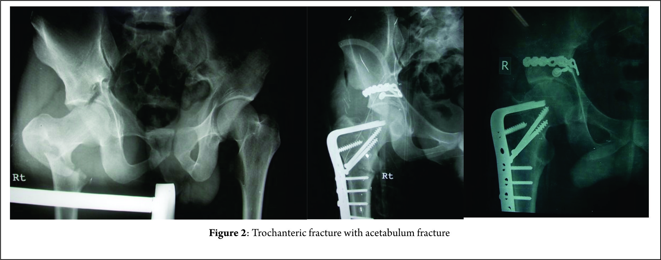
Floating hips have been classified as per Liebergall into groups A and B (5). Group A included femoral fractures with an ipsilateral unstable vertical shear or open book pelvic fracture [Fig 1]. In Group B fractures of the acetabulum were concomitantly present with a femur fracture. Technically only the latter would classify as a floating hip [Fig. 2].
Mueller classified floating hips into 3 types (4). Type A, a combination of acetabular and femoral fractures, type B combination of pelvic and femoral fractures, while type C was a combination of fractures of acetabulum pelvis and femur.

While the above 2 classification were based on the location of the fractures, later in 2002, in another article Liebergall proposed two types of injury and correlated it with its mechanism of action (3). The first is a posterior type injury: a posterior type acetabular fracture with ipsilateral diaphyseal femoral fracture. This was said to be a consequence of Dashboard injury where there was a direct blow to the knee. The force was transmitted from the femur to the posterior elements of acetabulum as the hip was in a flexed position. This caused a posterior wall or a transverse with posterior wall fracture of the acetabulum. For this combination to occur the acetabular fracture must have preceded the femur fracture following which there was still persistent force to cause bending forces in the diaphysis and cause a short oblique or a transverse fracture. These fracture patterns can also be associated with ligamentous derangement of the knee (tear in PCL) and or patella fracture with or without knee effusion (3).
The second type was the central type wherein there was a central type acetabular fracture and an ipsilateral proximal femoral fracture. The central fracture dislocation is an old eponym to describe the inward protrusion of femoral head into the pelvis which occurs unceasingly due to a bi-columnar fracture or displaced anterior and transverse fractures without posterior involvement. This pattern of injury was found in patients who have a history of fall and among pedestrians struck by a moving four-wheeler. Here the impact of injury was the lateral aspect of the greater trochanter and the transfer of force was to the hip joint which was transmitted centrally across the hip into the pelvis. The remaining force dissipated to the surrounding structures causing a fracture in the proximal femur if it hadn’t occurred at the time of impact. There were no associated knee injuries in these patients.
This suggested mechanism of injury and its subsequent consequences have been disputed by a retrospective study on 57 patients by Burd et al (9). Though data on mechanism of injury were not available in their study, there were no significant correlation between the type of femur fracture and associated acetabular fractures as described by Liebergall (9).
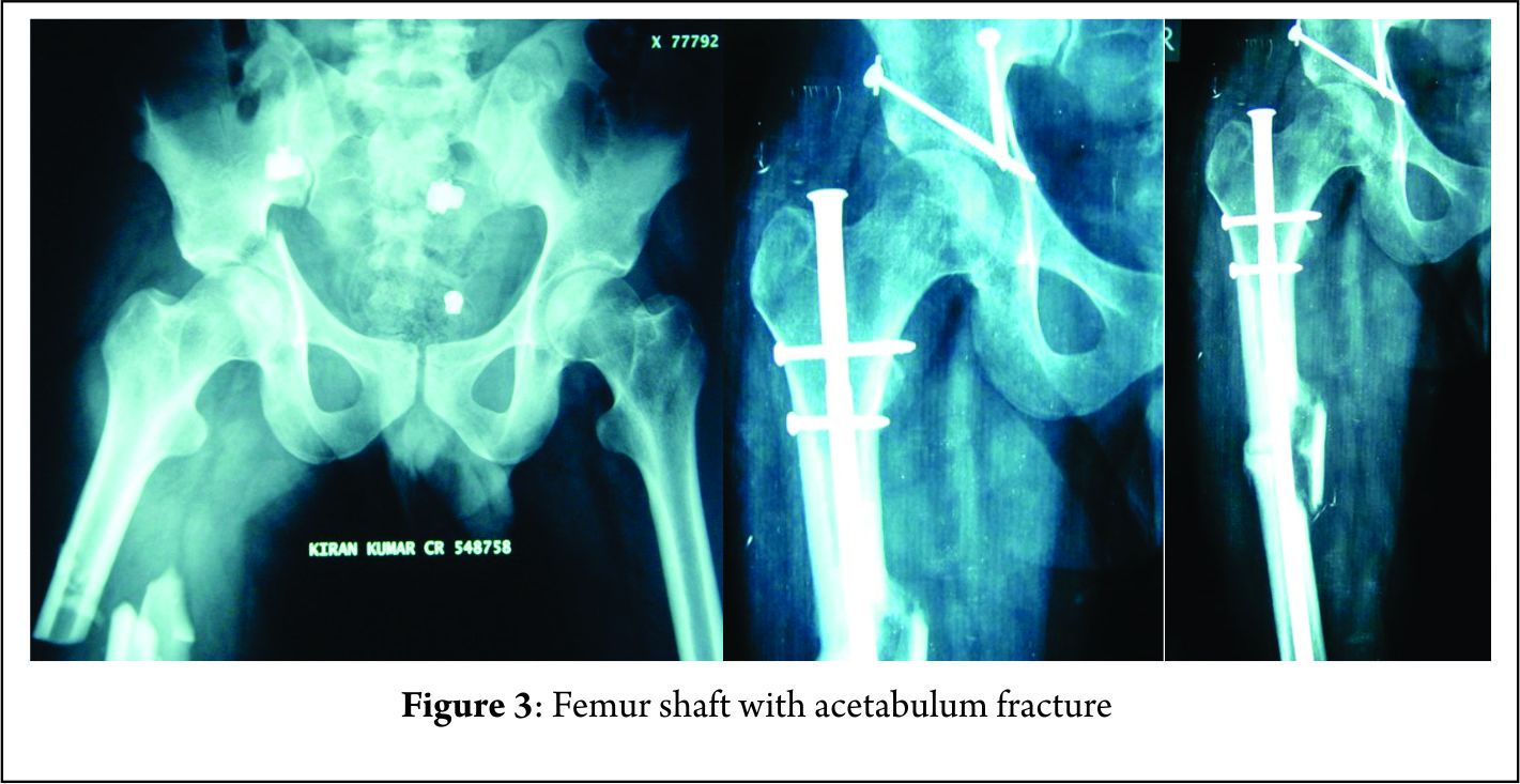

Femur fractures are mostly trochanteric or diaphyseal [Fig.2,3] rarely involving the neck of femur while distal femur fractures were not reported in the series of Liebergall (3, 5). He opined that the distal femur absorbs most of the energy because of its proximity to the point of impact and the residual force that is applied to the acetabulum is insufficient to induce a fracture. Despite the mechanism being a Dashboard injury with axial loading forces there is low prevalence of distal femur fractures. Suzuki et al reported four distal femur fractures in a total of 34 cases, while Burd et al in their retrospective study of 57 patients found 18 cases of distal femur fracture (9, 12). Most of the fractures of the femur are closed fractures while open femur fractures were found to occur in 22% of the cases in the study by Wu et al (15).

The patterns of acetabular fractures in cases of floating hips mostly are of the posterior wall or transverse fractures or their combination (9). Other elemental and associated fractures are possible and are found to be present sporadically. The natures of these fractures are a result of force transmission along the long axis of the shaft of or along the axis of the femoral neck. The various fracture patterns are possible due to the varying position of the involved limb in terms of rotation and or abduction and adduction of the affected hip. Fractures of the pelvis have not shown a particular trend to be universally applicable to floating hips. If the stable fractures Tile A are to be excluded then most of the fractures are found to be rotationally unstable as compared to both vertically and rotationally unstable (9,15). The commonly found injuries of the pelvis are the lateral compression injuries as described by Suzuki et al and the Tile B type of pelvis fracture as described by Wu et al constituting 65 % and 75%of the total injuries respectively ( 12, 15). Liebergall et al in their study had encountered 17 cases of which 9 were vertical shear while 3 were vertical shear combined with open book type injury (5).

Management

As with any case, poly trauma management proceeds in line with ATLS protocol. Patients should be assessed for other associated injuries which may require lifesaving surgeries involving other systems. Embolization should be considered in patients with major pelvic fractures who are haemodynamically unstable, have evidence of pseudo aneurysm or cases where blood pressure does not respond to massive transfusion (16). The time to first definitive surgery irrespective of fixation of femur or acetabulum was found to be 87 hours and 132 hours as shown by Burd et al and Muller et al respectively (9,4). Thus, showing the necessity of adequate resuscitation and adherence to principles of damage control orthopaedics. Early fixation of fracture plays a crucial role in decreasing the chances of neurovascular and pulmonary complications.
The controversy arises when we consider the order of fractures to be fixed. There have been mainly two views put forth, Liebergall (5) classically said that the femur fracture fixation should take precedence over acetabular fracture which was also followed by Suzuki et al (12) and suggested by Kregor and Templeman (17), while Muller (4) operated femur first in only 38 per cent of his cases. Immediate stabilization of the pelvic injury in the emergency room as a resuscitation tool was to be followed by fixation of the femur. In cases with a concomitant acetabular fracture Muller et al stated that floating hips do not represent special treatment and can be treated as per existing guidelines for the aforementioned fractures (4).
Liebergall stated in his article the hypothetical need for two operating surgical teams taking into consideration the polytrauma presentation of these injuries. He advocated early stabilisation of the femur fracture as per prevailing guidelines and a three to five-day delay for the fixation of acetabular fractures (18). Femur fracture was fixed first followed by fixation of unstable vertical shear injuries. SI joint disruptions were approached via 2 curvilinear incisions. Reduction of the fracture was followed by posterior stabilisation with Harrington sacral compression rods. Sacral ala fractures were reduced and stabilised with interfragmentary compression screws. None of the cases quoted in the study required anterior fixation of pelvic injuries (3). Acetabulum fractures which involved the dome and posterior wall or column were all approached with the same Kocher-Langenbeck approach. There was need for anterior Ilio-femoral approach and a trochanteric osteotomy when fracture lines extended up to the iliac wing or the anterior column. In cases where intramedullary nailing was to be done trochanteric osteotomy was done first followed by nailing and then the acetabulum was reduced and fixed. The surgery concluded with fixation of the trochanteric osteotomy. Earlier fixation of the femur also facilitates easier positioning, preparing and draping of the patient (9). In fractures of the femur involving the diaphysis or the distal third, fixation with plates and distal femur nails are preferred. This is desirable as we can avoid incisions proximally and preserve the anatomy of the area around the acetabulum. Proximal femur fractures can be approached laterally in the supine position on a fracture table and be fixed with a sliding hip screw and blade plates. The fixation of the acetabulum can proceed after transfer to a radiolucent table. If antegrade intramedullary nailing is required, it should be done preferably in the lateral position to incorporate its incision with the Kocher-Langenbeck incision. Either the piriformis or the greater trochanter can be used as the entry point. But the problem occurs while trying to visualise the femoral heads lateral projection to check the direction of the proximal locking blade or screw. The advantage here is that there is no need to change positions for fixation of acetabular fractures.
Beginning in the supine position facilitates monitoring of patient vitals and treatment of other abdominal, thoracic and associated injuries in other limbs. Suzuki et al and Wu et al in his paper on this topic fixed unstable pelvic fractures in the emergency room with external fixation (12, 15). Suzuki et al in their paper performed internal fixation of pelvis after femur fixation in 6 patients while in 3 patients pelvis was fixed before femur in the same anaesthesia. In a case with minimally displaced acetabulum fracture they broke their protocol to fix the femur first in fear of displacement of the acetabulum fracture (12).
Kregor and Templeman in their paper suggested three different strategies for fixation of floating hips: Fixation of the acetabulum followed by antegrade nailing, or fixation of acetabulum followed by plating of femur, and finally distal femur nailing of femur followed by acetabular fixation. He opined early preference for the acetabular fracture in views of preventing further damage to the hip joint (17).
Cases where a concomitant dislocation is present along with the floating hip, authors primarily focussed on the reduction of the dislocated hip [Fig.4] (19, 20). Tiedeken et al in his case opened the hip by the Kocher-Langenbeck approach and the joint was reduced and posterior wall acetabulum plated with reconstruction plates. The patient was then placed in supine position to fix the femur fracture by the retrograde method (19). Duygulu et al in their paper approached the hip by the posterolateral approach and fixed the transverse and posterior wall fractures by reconstruction plates. In this case there was an associated neck of femur fracture along with shaft, a reconstruction nail with a piriformis entry was used. Closed reduction was done of the shaft and the nail driven through. This was followed by the reduction of the femoral head. An external fixation for pubic diastasis was applied (20).
In case reports of a concomitant floating hip and knee injury the authors have chosen to fix the tibia first followed by the femur and then finally check the stability of the acetabulum and proceed. Both the long bones were fixed via the same incision (21). Hideto et al in their case report of an ipsilateral femoral neck, shaft and acetabular fractures, the femur shaft was fixed in the same sitting as the neck fracture. The shaft femur fracture was fixed by retrograde nailing and then patient was transferred to a traction table where the neck of femur fracture was fixed with multiple cannulated screws. The acetabulum fracture was operated on seven days after the femur surgery. The transverse acetabulum fracture was approached by the modified Stoppa technique and fixed with reconstruction plate. This was followed by posterior wall fixation by the Kocher-Langenbeck approach (22).

Complications

In a metaanalysis by Giannoudis et al acetabular fractures were found to have complication rates as described below. Traumatic and Iatrogenic nerve injuries were found to be present in 16.4% and 8 % respectively. Other complications documented were DVT/PE, local infection, heterotopic ossification, and AVN which were found to be present in 4.3%, 4.4%, 5.7% and 5.6%respectively. The most commonly found complication was osteoarthritis of the hip occurring in 19.8% of the cases (23).
The rates of complications reported by authors in patients of floating hips vary widely. However there seems to be an increase incidence of complications seen in cases of floating hips when compared to those of isolated acetabular fractures. Leibergall in his study of 17 hips encountered two cases of delayed wound healing. There were three cases of pin tract infection which healed by removal of the fixator. 4 patients had severe pulmonary and or cardiac complication with one requiring a tracheostomy. Serious long term morbidity was present in 10 of the seventeen cases, two of which were iatrogenic sciatic nerve paresis. Other complications included no anatomic acetabular dome, heterotopic association, painful heel, peroneal and femoral paresis and subtalar osteoarthritis. One patient had a reflex sympathetic dystrophy of the foot while one case needed an above knee amputation. 5 patients had shortening up to 2.5 cm of the involved extremity. The shortening was due to comminution in the femoral fracture site in four of these cases, while the remaining one was attributed to the malunion of pelvis (5).
The complications documented by Suzuki et al in their paper of floating hip were 5 cases of neurological injuries and one case each of fat embolism, non-union, and displacement of pubis, heterotopic ossification, aseptic necrosis and one deep wound infection. Fat embolism was present on the first day following the injury. Deep infection occurred in the case which had presented with the pelvic ring fracture and a Morel – Lavallee lesion. Of the five Neurological injuries, 3 were present at presentation and two cases operated by the ilioinguinal approach developed lateral cutaneous nerve palsy post operatively. Class III bookers heterotopic ossification occurred in one patient which was incidentally operated via a single incision for both femoral shaft fixation and acetabular surgery. (5)
Burd et al in their study had documented complications of deep vein thrombosis in seven patients (12%), non-union of the femur in two patients (3%) and avascular necrosis of the femoral head in one patient (2%). Post traumatic osteoarthritis of the involved hip joint was seen in nine patients (16%). Heterotopic ossification (34%) was classified by the Brooker classification where 4 cases were clinically not significant while the remaining 7 cases had lesions of Brooker grade 3 or above. Nineteen of the fifty seven (33%) cases had evidence of Sciatic nerve injury. Seven of these patients had sensory involvement, five had motor involvement while the remaining seven had involvement of both. It was noted that at an average of seven and half months post operatively eight cases had recovered completely four of which were of the sensory type and two each were the motor and combined types respectively. Two other cases showed partial recovery at eleven and half months post operatively while the remaining nine cases showed no improvement till a little above seventeen months of follow up. Trendelenburg gait was found to be present in eight patients (14%) in the post-operative period (9).
Mueller in his study had a 35 % occurrence of sciatic nerve injury. The recovery rate established for his sciatic nerve injuries were 25%. There was one case of vascular injury reported (4).
In an article by Zamora et al of 11 floating hips acetabular fractures were most commonly associated with diaphyseal fractures. 3 of the cases were supracondylar level fractures and were associated with popliteal artery injury. Despite prompt vascular repair the limbs were not salvable and had to undergo an above knee amputations. In their series, this was the most dreaded complication (24).

Conclusion

Floating hips are a wide spectrum of injury patterns which require surgical intervention. Fractures involving the pelvis take preference over any other orthopaedic injury which may be managed according to existing protocols. Cases involving unstable pelvic fractures have been fixed by external fixation equivocally and the resuscitation of the patient takes precedence before definitive fixation. Controversy arises as to how we proceed in view of order of fixation in a case of floating hip. Proponents of earlier fixation of femur suggest factors such as ease of fixation of acetabulum as traction application is easier, easier preparing draping and positioning for acetabular fixation and reduction in risk of fat embolism. In other cases, where retrospective data has been used the order of fixation has been left upon the discretion of the trauma surgeon (12). However, earlier fixation of the acetabulum has been propagated in cases of unstable dislocated hips and irreducible dislocated hips. Antegrade nailing in these cases could compromise later acetabular surgery by distorting and de vascularizing the soft tissue and musculature. Antegrade nailing is also thought to carry risk of avascular necrosis and weakness of the hip abductors as described by Kregor et al (17). Proponents of femur fixation first also had to abandon their protocol in fear of gross displacement of the acetabular fractures in undisplaced cases (12).
The surgical order in management of floating hips has been up for debate. No definitive guidelines are available for the order of fixation of these fractures. This is our attempt to give a structured approach for the management if floating hips.
Unstable Fractures of pelvis are mostly of the lateral compression type of Young Burgess Classification or type B type of the Tiles classification which can be fixed by the use of external fixation preferably in the emergency room. Our focus now shifts to the hip joint, to ascertain it is reduced or not. In cases of irreducible dislocated hips open reduction is recommended before fixation of femur. In cases of central type of floating hip the femur can be fixed first either by means of distal femur nail or distal femur locking plate. This will facilitate traction and aid in fixation of acetabulum and other pelvic fractures later on. For fractures of the proximal femur antegrade nailing or dynamic hip screw as ascertained by the fracture type are preferred. The incision of antegrade nailing can be incorporated into the Kocher- Langenbach approach if such an approach is desired. Neck of femur fractures are to be given priority of fixation before fixing fractures of the femur or at the same sitting. The acetabular and other fractures of the pelvis like sacrum fractures, SI joint disruptions can be fixed at a later stage.


References

1. Agarwal A, Chadha M. Floating injuries: a review of the literature and proposal for a universal classification. Acta Orthop Belg. 2004; 70: 509–14.1. Agarwal A, Chadha M. Floating injuries: a review of the literature and proposal for a universal classification. Acta Orthop Belg. 2004; 70: 509–14.2. Simpson NS, Jupiter JB. Complex fracture patterns of the upper extremity. Clin Orthop Relat Res. 1995; 318: 43–53.3. Liebergall M, Mosheiff R, Safran O, Peyser A, Segal D. The floating hip injury: patterns of injury. Injury. 2002; 33: 717–22.4. Muller EJ, Siebenrock K, Ekkernkamp A, Ganz R, Muhr G. Ipsilateral fractures of the pelvis and the femur–floating hip? A retrospective analysis of 42 cases. Arch Orthop Trauma Surg. 1999; 119: 179–82.5. Liebergall M, Lowe J, Whitelaw GP, Wetzler MJ, Segal D. The floating hip. Ipsilateral pelvic and femoral fractures. J Bone Joint Surg Br. 1992; 74: 93–100.6. Wiltberger BR, Mitchell CL, Hedrick DW. Fracture of the femoral shaft complicated by hip dislocation—a method of treatment. J Bone Joint Surg Am 1948; 30A: 225–28. 7. Chi-Chuan W, Chun-Hsuing S, Lih-Huei C. Femoral shaft fractures complicated by fracture-dislocations of the ipsilateral hip. J Trauma 1993; 34: 70–5.8. Helal B, Skevis X. Unrecognized dislocation of the hip in fractures of the femoral shaft. J Bone Joint Surg Br 1967; 49B: 296–300.9. Burd T, Hughes M, Anglen J. The floating hip: complications and outcomes. J Trauma 2008; 64: 442–8. 10. Zamora-Navas P, Guerado E. Vascular complications in floating hip. Hip Int. 2010; 20: S11–8. 11. Iotov A, Tzachev N, Enchev D, Baltov A. Operative treatment of the floating hip. J Bone Joint Surg Br 2006; 88-B(Supp I): 160. 12. Suzuki T, Shindo M, Soma K. The floating hip injury: which should we fix first? Eur J Orthop Surg Traumatol 2006; 16: 214–813. Willett K, Al‑Khateeb H, Kotnis R, Bouamra O, Lecky F. Risk of mortality: The relationship with associated injuries and fracture treatment methods in patients with unilateral or bilateral femoral shaft fracture. J Trauma 2010; 69: 405‑1014. GiordanoI V, Amaral NP, Rios H,Franklin CE, Pallottino A. Management of ipsilateral fractures of the femur and pelvis (floating hip): a prospective study on 16 cases Rev bras ortop. 2007; 42: 9 15. Wu CL, Tseng IC, Huang JW, Yu YH, Su CY, Wu CC. Unstable pelvic fractures associated with Femoral shaft fractures: a retrospective analysis. Biomed J. 2013; 36(2): 77-83.16. Panetta T, Sclafani SJ, Goldstein AS, Phillips TF, Shaftan GW. Percutaneous transcatheter embolization for massive bleeding from pelvic fractures. J Trauma 1985; 25: 1021- 917. Kregor PJ, Templeman D Associated injuries complicating the management of acetabular fractures. Orthop Clin North Am 2002; 33: 73–9518. Tile M. Fractures of pelvis and acetabulum.2nd ed. Williams, Baltimore 1995 [ chapter 19]19. Tiedeken NC, Saldanha V, Handal J, Raphael J. The irreducible floating hip: a unique presentation of a rare injury.  J Surg Case Rep. 2013 Oct; 2013(10):20. Duygulu F, Calis M, Argun M, Guney A.Unusual Combination of Femoral Head Dislocation Associated Acetabular Fracture With Ipsilateral Neck and Shaft Fractures: A Case Rreport. J Trauma. 2006;61:1545–154821. Yashavantha C, Nalini K B, Nagaraj P, Jawal A. Ipsilateral Floating Hip and Floating Knee – A Rare Entity Journal of Orthopaedic Case Reports 2013 July-Sep;3(3):  3-622. Irifune H, Hirayama S, Takagi N, Narimatsu E. Ipsilateral Acetabular and Femoral Neck and shaft fractures. Case Rep Ortop. 2015; 2015 : 35146523. Giannoudis PV, Grotz MR, Papakostidis C, DInopoulus H. Operative treatment of displaced fractures of the acetabulum. A metaanalysis. J. Bone Joint Surg Br. 2005 Jan; 87(1): 2 -9 24. Zamora -Navas P, Guerado E. Vascular complications in floating hip.Hip Int. 2010;20  7:S11-8.


How to Cite this article: Sen R, Jha L.Floating Hip. Journal of Clinical Orthopaedics Jan – June 2017; 2(1):43-48.

(Abstract    Full Text HTML)      (Download PDF)


Spinal tuberculosis – an Update

Vol 2 | Issue 1 |  Jan – June 2017 | Page 31-42 | Kshitij Chaudhary, Arjun Dhawale, Ram Chaddha, Vinod Laheri


Authors: Kshitij Chaudhary [1], Arjun Dhawale [1], Ram Chaddha [2], Vinod Laheri [2].

[1] Sir HN Reliance Foundation Hospital, Department of Orthopaedics and Spine Surgery; Mumbai, India
[2] Apollo Hospital, Navi Mumbai, Maharashtra. India

Address of Correspondence
Dr. Kshitij Chaudhary
Sir HN Reliance Foundation Hospital, Department of Orthopaedics and Spine Surgery; Mumbai, India
Email: chaudhary.kc@gmail.com


Abstract

Musculoskeletal tuberculosis, especially spinal tuberculosis is a challenging scenario. The disease presentation varies and profile of the organism is changing rapidly with rise of antibiotic resistance. The management protocols need to be revisited in light of new information and current review is aimed at achieving this goal. This review article is a summary of the symposium conducted by Bombay Orthopaedic Society at KEM Hospital in November 2016. The focus of this discussion was primarily on nonoperative management of spinal tuberculosis including focus on diagnostic protocol and medical management controversies. Surgical aspect of the disease is also covered with recent advances in the spine surgical protocols mentioned in brief.
Keywords: Spinal Tuberculosis, diagnosis, antibiotic resistance, surgery.


1. Introduction

Spinal tuberculosis is the most common infection affecting the skeletal system in our country, and if incorrectly treated, can have devastating and permanent sequelae. The emergence of drug-resistant tuberculosis and HIV has turned this age old infection into a deadly and terrifying disease. Poor socioeconomic conditions and an unhygienic living environment contribute to the persistence of the infection in our society. Poor access to quality healthcare and indiscriminate and unscientific use of second-line antibiotics has stoked the fire as we stand and stare helplessly at the impending epidemiological disaster. It behooves us as representatives of the medical community to educate ourselves and others regarding this problem and practice recommended treatment guidelines.

2. Clinical Features

The most common presenting symptom of spinal tuberculosis is back pain or neck pain. Most patients seek consultation after weeks or months of pain as the onset is usually insidious and progression is slow. Frequently it is mistaken as benign low back pain, and treated with painkillers as the early radiographs are often normal. Patients may experience constitutional symptoms (reported incidence varies between 17-54%), however, compared to pyogenic spondylodiscitis fever, anorexia, fatigue are less common. Therefore, the clinician should maintain a high degree of suspicion, especially in patients who complain of ongoing pain for more than a month, have rest pain, or if associated constitutional symptoms are present. In addition, as tuberculosis affects the anterior column primarily, a knuckle deformity, i.e. prominence of a single spinous process might be one of the early signs of tuberculous spondylodiscitis. Some patient can also present with a cold abscess in remote locations like the posterior triangle of the neck (cervical spine TB), along the ribs (thoracic spine TB), or in the inguinal region (lumbar spine TB). The clinician should also maintain a high degree of suspicion in immunocompromised patients, especially HIV infections, and should also be wary of patients with a history of tuberculosis, or those who have come in contact with tuberculosis patients.Unfortunately, in our country, many patients are diagnosed late and present with complications that may require surgical intervention. There are two main complications of spinal tuberculosis – spinal deformity and neurological deficit. The spinal deformity that typically develops as a consequence of anterior spinal column destruction is kyphosis. The kyphosis is usually of an angular variety that can cause long-term consequences if severe. The most grave among them is late-onset myelopathy due to a stretch of the spinal cord over an internal gibbus. In children, kyphosis can continue to progress after healing. The children who are more likely to have progressive deformity are identified using the “spine at risk” signs as described by Rajasekaran. These radiographic signs are seen when the posterior elements become incompetent, thus resulting in progressive deformity.Neurological deficits are mainly seen in cervical and thoracic tuberculosis. The cause of neurological deficit can be “soft” or “hard” compression (described in detail in the section on radiological features). Another rare cause of neurological deficit is inflammatory vascular thrombosis of spinal arteries resulting in spinal infarct. There are no diagnostic imaging findings to diagnose a spinal infarct, and this is usually a diagnosis of exclusion.

Radiographic Features

3.1.  Radiographs

Standard radiographs, anteroposterior and lateral views, are used commonly as the first line of investigation; however, they suffer from several disadvantages. Radiographic changes are usually not apparent until more than 50% of the vertebral body is destroyed, thus delaying the diagnosis. Junctional areas and posterior elements are difficult to visualize. Disc space narrowing with endplate erosions (paradiscal lesions) are early radiographic signs. With progressing and destruction of the spinal column, the radiographs may show varying degree of focal kyphosis due to vertebral body collapse. A paraspinal abscess may cast soft tissue shadow, which in the thoracic spine may be difficult to differentiate from the descending aorta. Abscess in cervical spine will show up as an increase in retropharyngeal space and those in the lumbar spine may result in asymmetry or bulging of the psoas outline. In chronic cases, calcifications in abscess wall are pathognomonic of tuberculosis. Calcifications are formed because, unlike pyogenic bacteria, MTB lacks proteolytic enzymes. Progression of kyphosis or vertebral body destruction early in the course of chemotherapy (first three months) should not be considered as a sign of treatment failure.

3.2.  MRI

MRI is the imaging modality of choice for spinal TB and can detect infection early in its course. Paradiscal involvement is the most common type of lesion. Central body, subligamentous, posterior element tuberculosis can also sometimes be encountered. The entire spine should be screened as noncontiguous lesion are seen in 16-71% of patients. The recommended protocol for imaging is presented in Table 1.

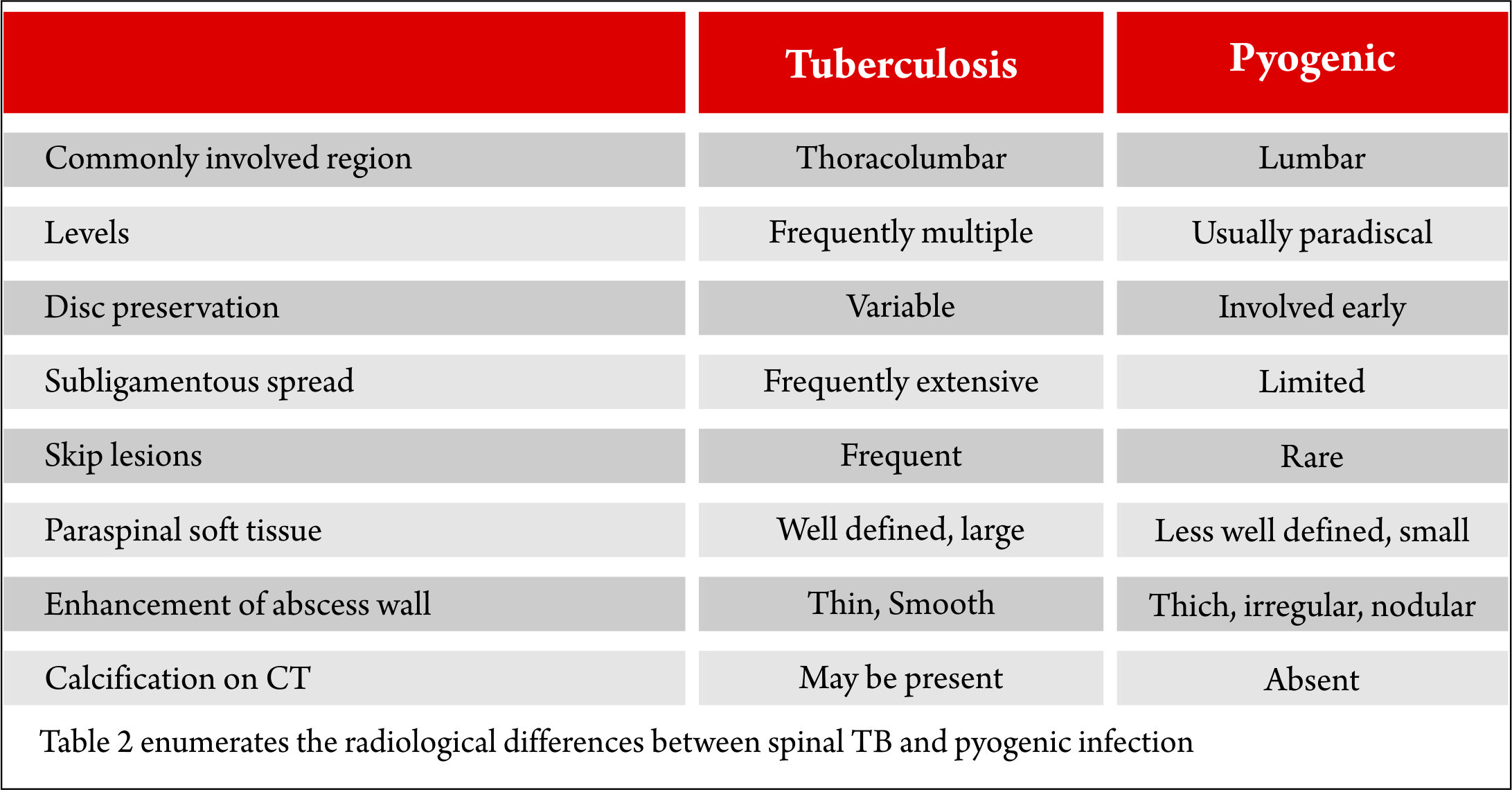
In many parts of the country, the diagnosis of spinal TB rests on imaging findings on MRI. In an endemic country like ours, the clinician usually is right because the odds are in his favor. However, there are no pathognomonic imaging features of spinal TB that can reliably differentiate it from other spinal infection or tumors. Hence, tissue diagnosis is mandatory and recommended. Table 2 enumerates the radiological differences between spinal TB and pyogenic infection; however, it is important to remember that these have a poor predictive value.


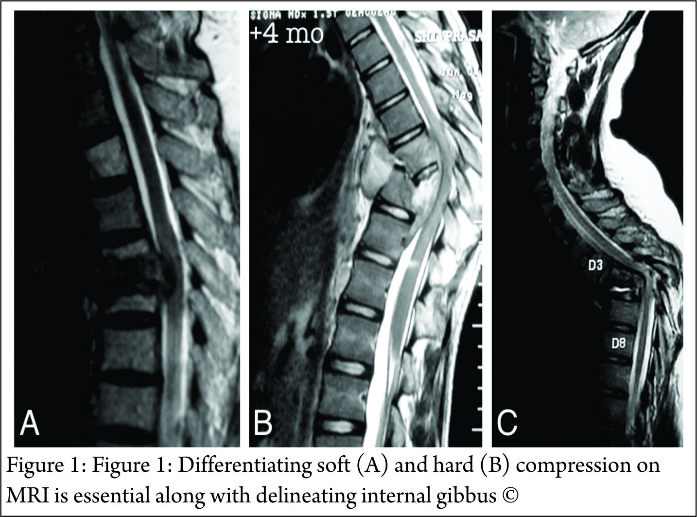
Brucellosis frequently affects the lumbar spine. Anterior osteophytes (parrot beak) is a typical radiological feature on radiographs. Intradiscal air is identified in about one-third cases. Fungal osteomyelitis is difficult to differentiate from spinal TB on imaging. Metastasis or spinal tumors frequently affect the pedicle and spare the disc. The paravertebral soft tissue involved in lymphomas demonstrates a low signal on T2 MRI. However, noncaseating or granular varieties of spinal TB may look similar. Early infection and type 1 Modic type degenerative changes may look similar, although a high signal in the disc should raise the suspicion of infective spondylodiscitis.MRI is useful for another purpose – to diagnose the type, extent, and severity of epidural spinal cord compression (Figure 1).

“Soft” compression should be differentiated from “hard” compressions as this has implications for the management of the patient. Abscess (diffuse hyperintense T2 signal and hypointense T1 signal) and caseous granulation tissue (heterogeneous hyperintense to isointense T2 signal) are “soft” compressions. Bony sequestrum and retropulsed disc (hypointense T1 and T2 signal) are “hard” compressions (Figure 1). Translations and internal gibbous causing spinal cord compression is another example of “hard” compression. “Hard” compressions that cause neurological deficit cannot be treated with chemotherapy alone and usually require surgical decompression. Rarely, non-compressive lesions, such as vascular infarct or meningeal inflammation) are the cause of neurological deficit. It is extremely difficult to prove these as the cause of spinal cord dysfunction, especially in patients with epidural spinal cord compression.

3.3.  CT scan

CT scan is primarily used to assess the extent of osseous destruction accurately before surgical intervention, especially in extensive infection. In some situations, it may help differentiate “hard” from “soft” epidural compression.

4. Biopsy

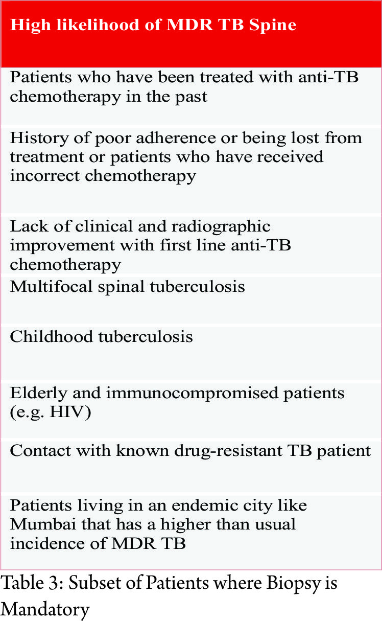
As we have seen, there are no pathognomonic imaging signs that can reliably differentiate spinal tuberculosis from other spinal infections or tumors. Hence, a biopsy to obtain tissue for histopathological and microbiological diagnosis is mandatory to confirm the diagnosis of spinal tuberculosis. Besides, the mounting incidence of multi-drug resistant tuberculosis has made biopsy unavoidable. The yield of spinal biopsy is variable and depends not only on the expertise of the surgeon or radiologist doing the biopsy but also on the availability of specialized, well-equipped laboratories and microbiologists. Access to such facilities is non-existent in resource-poor regions of our country. However, there is a particular subset of spinal tuberculosis patients in whom biopsy is mandatory as in these patients the probability of encountering drug resistance is high (Table 3).


In patients with uncomplicated spinal TB (i.e. patients who are not planned for surgical intervention), a closed core biopsy is indicated. CT-guided core biopsy is ideal to acquire a representative sample safely, however, when unavailable, a fluoroscopy-guided biopsy can work as well, especially if the target area is easily accessible. Core biopsy (10-14G bone biopsy needle) is preferred to fine needle aspiration cytology or FNAC (18-22G needles) as the diagnostic yield of the latter is poorer. In any case, aspiration biopsy if required can always be performed via a core biopsy needle. Higher microbiological yield is obtained if paraspinal or prevertebral abscesses are targeted for pus sample rather than bone cores. If the patient has multiple sites of spinal TB, a more easily accessible and safer area is chosen. The biopsy can be performed via a transpedicular or extrapedicular approach, and the choice depends on the target area of biopsy. Anterolateral approach is required in cases of cervical spine TB. The neuromuscular bundle can be displaced manually, and the needle can be placed under CT image guidance. This technique requires considerable expertise and a confident radiologist. Biopsy for craniovertebral tuberculosis can be performed either transorally or via a posterolateral approach. Microbiological samples are collected in sterile containers in saline (2-3ml is enough for the purpose is only to keep the tissue hydrated; if a large quantity of saline is used the lab has to centrifuge to isolate the tissue) and sent immediately to the lab. Histopathological samples are collected in 10% formalin. The following tests should be ordered on the samples (Table 4).

4.1.  MGIT Cultures and Drug sensitivity testing

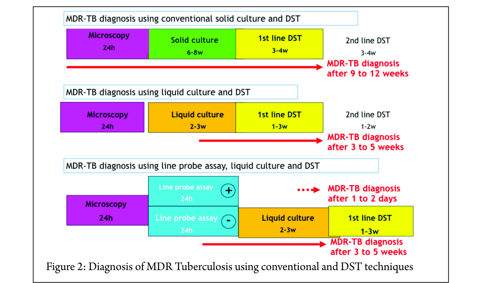
Definitive diagnosis of Mycobacterium tuberculosis infection rests on positive microbiological culture from the tissue sample. The traditional method of culture using Lowenstein-Jensen solid medium has been replaced with BACTEC™ MGIT™ liquid cultures. MGIT cultures are more sensitive (50 to 60% in osteoarticular tuberculosis) and can give faster results. Results are available in a few weeks (maximum 45 days) (Figure 2).

Prior treatment with chemotherapy can lower the yield and hence when possible it is better to perform a biopsy before initiating chemotherapy. It is not recommended to stop chemotherapy if the patient is on antitubercular medications only for the purpose of biopsy. If the results are positive, one should ask for phenotypic drug sensitivity testing (DST) for first-line drugs. In addition, phenotypic DST can be done for second-line drugs if one suspects MDR tuberculosis. The approximate cost of BACTEC™ MGIT™ liquid cultures is Rs. 1000-1500. First-line DST costs about Rs. 4000-5000 and second-line DST costs about Rs. 5000-6000.Most labs will also perform direct smear and AFB staining before putting the sample in for culture. The sensitivity of direct smears is poor (less than 10%) in osteoarticular TB cannot be relied on to make a diagnosis.

4.2.  Histopathological examination (HPE)

Histopathological diagnosis is usually obtained in about 60% of spinal TB patients. The classical histopathological features are caseating necrosis, epitheliod cell granuloma, lymphocytic infiltrate, and Langhans giant cells. It is important to remember that granulomas can be detected in other infections and inflammatory disorders as well. Therefore a positive HPE diagnosis can only suggest a probable diagnosis of mycobacterial infection. AFB staining of HPE samples can sometimes detect mycobacteria (not necessarily Mycobacterium tuberculosis), but as with direct smear, the sensitivity is quite low.

4.3.  Other cultures

It is a good custom to always ask for pyogenic bacterial cultures as well. This practice may detect a primary pyogenic infection or rarely secondary bacterial infection in addition to tuberculosis. It is also important to consider the possibility of fungal infections in susceptible and at-risk patients.

4.4.  GeneXpert

GeneXpert is a new molecular test that detects the presence of Mycobacterium tuberculosis DNA. The main advantage is its rapid turnaround time. Results are usually available within 6 hours. It can detect Rifampicin resistance as well and thus aid in starting MDR treatment early in the course of therapy. As it is a DNA-based test, the specificity is 100%. However, the sensitivity is quite low of osteoarticular tuberculosis (about 60 to 70%). Therefore a negative GeneXpert cannot be used to rule out tuberculosis infection. The test is not widely available, especially in remote areas of the country. However, wherever feasible, it can be a useful adjunct to the above tests. The approximate cost is about Rs. 1500-2000 and in many centers, it is available free of charge through public-private partnerships under the DOTS program.

4.5.  Line Probe Assay

Line probe assay or LPA is another DNA PCR-based molecular test that is specifically used for detecting drug resistance. The first-line kit detects Isoniazid and Rifampicin resistance and aids in the diagnosis of MDR-TB. The second-line kit detects Ethambutol, cyclopeptides (Capreomycin, Kanamycin, Amikacin and Viomycin) and fluoroquinolone resistance and aids in the diagnosis of XDR-TB. WHO recommends its use for smear-positive respiratory samples only. However, this test can be used as an alternative to phenotypic DST after MGIT cultures are positive. The results are available within 48 hrs (compared to phenotypic DST which requires about 3 weeks) and a diagnosis of MDR or XDR can be established (Figure 2). The information provided by this test can streamline MDR or XDR therapy earlier in the course of therapy. It is not recommended to rely only on LPA results as monoresistance to drugs not included in the kit can be missed. Therefore, phenotypic DST is always required to complete drug sensitivity testing. If the patient has a low risk for drug resistance, then phenotypic DST is adequate. If the patient has a high risk for drug resistance, then LPA followed by phenotypic DST is advocated if the clinician wants to establish the diagnosis of MDR or XDR to guide therapy. The main disadvantage is the cost. First-line kits cost Rs 2000-3000 and second-line kits cost about Rs. 5000-60005.

Other investigations

Other investigationsCBC, ESR, CRP are a measure of disease activity and are frequently used to monitor therapeutic response to chemotherapy. They are more likely to be markedly abnormal in pyogenic infection compared to tuberculosis. Xray Chest should be done, as up to 67% patients may have either active focus or healed sequelae of pulmonary tuberculosis. Tests to detect HIV should be performed in high-risk patients or patients presenting with extensive or atypical spinal TB. Serological tests (IgM, IgG titers) and interferon release assays (Quantiferon TB Gold, TB-SPOT test) cannot differentiate latent from active infection and are not recommend by WHO. Mantoux test is of limited value in an endemic country like India. LFT and RFT are ordered as a baseline before starting chemotherapy and later to monitor side-effects.

6.  First line chemotherapy

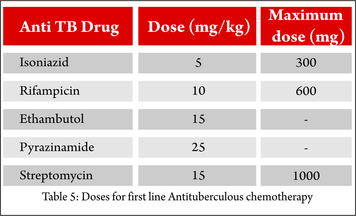
As with pulmonary tuberculosis, multidrug chemotherapy is the mainstay of treatment for spinal tuberculosis. Empirical treatment is usually started after biopsy before confirmation of mycobacterial infection. It is advisable to involve a chest physician or infectious disease specialist early in the course of therapy. The drugs that comprise first-line chemotherapy are Isoniazid (H), Rifampicin (R), Pyrazinamide (Z), and Ethambutol (E). The WHO recommends two months of intensive chemotherapy comprising of HRZE followed by a continuation phase of HR. The doses are weight-dependent (Table 6)

and hence fixed dose combinations should be avoided (e.g. Tab AKT 4 kit has 450mg Rifampicin that is inadequate for more than 60kg individual). There is no evidence to show that corticosteroids improve or enhance treatment effect of antibiotic and may be detrimental. However, it is a common practice (although not evidence-based) to start a short course of steroids for patients presenting with an acute neurological deficit.

6.1.  Duration of multi-drug chemotherapy

There is a lack of consensus regarding the ideal duration of multidrug chemotherapy for spinal TB. WHO recommends nine months of treatment for TB of bones and joints (2HREZ + 7 HR) because of the serious risk of disability in addition to difficulties in assessing treatment response. British Thoracic Society (BTS) recommends six months (2HREZ + 4 HR) of chemotherapy. American Thoracic Society (ATS) recommends six months of chemotherapy in adults and 12 months in children for spinal TB. Inadequate or unessential, prolonged duration of treatment should be avoided.

6.2.  Role of DOTS (directly observed therapy short course)

Under the newer regime (National TB program), DOTS is no longer supportive of alternate day therapy, and daily treatment is recommended.

6.3.  Adverse effects of First-line anti-TB drugs

The common adverse effects are nausea, vomiting, hepatitis (HRZ), peripheral neuropathy (H), discolored body fluids (R), color/night blindness (E), joint pains (Z). It is best to consult the treating physician if the patient develops adverse effects as the drugs may need to be modified or discontinued.

6.4.  TB and HIV co-infection

The risk of developing tuberculosis (TB) is estimated to be between 26 and 31 times greater in people living with HIV (PLHIV) than among those without HIV infection. HIV-positive patients are more likely than HIV-negative patients to have extrapulmonary TB or smear-negative pulmonary TB. HIV testing is recommended in patients who were diagnosed with TB. The preferred recommendation for many TB-HIV patients is to start and complete TB treatment, and then start ART (antiretroviral therapy). However, if the patient’s clinical status is poor (other signs of HIV clinical stage 3 or 4 or CD4 count is less than 350/mm3), it may be necessary to refer the patient for ART treatment sooner. If patient is not on ART, start TB treatment immediately, or if already started, continue TB treatment.

6.5.  Judging treatment response to multidrug chemotherapy

The treatment response is mainly monitored using clinical and radiological evidence. Biochemical markers such as CBC, ESR, and CRP are not reliable markers to assess healing. Clinical response to healing is judged by resolution of constitutional symptoms, weight gain, improved appetite, reduction in spinal pain, and progressive increase in activity. Radiographs may show initial deterioration, however eventually healing is seen as remineralization of bone and sclerosis of vertebral bodies. The end result of healing may be spontaneous fusion or a stiff fibrous ankylosis. Routine interval MRIs on follow-up are not recommended, especially in the initial few months of antibiotic treatment. A paradoxical worsening of MRI findings can be noted up to 3 months, and this is believed to be secondary to an immunological response to the dying bacilli. MRI is indicated if there are reasons to suspect drug resistant or poor response to antibiotics. MRI findings of healing are a resolution of marrow edema with or without its conversion to a fatty marrow. An abscess may get walled off, and sterile collections may be encountered on MRI for years following the termination of treatment. Hence, by itself, abscess on MRI at the end of treatment is not taken as a sign of persistence of infection.If the clinical and radiological response is poor or inadequate, the surgeon must consider these possible scenarios: 1) drug resistance, 2) late responder, 3) mechanical or instability related pain, or 4) incorrect diagnosis of spinal TB. This may require a repeat biopsy or assessment of spinal column stability using dynamic or weight bearing radiographs. If the diagnosis is in doubt, in spite of following this protocol and the patient continues to deteriorate, a surgical debridement and column reconstruction may be indicated.

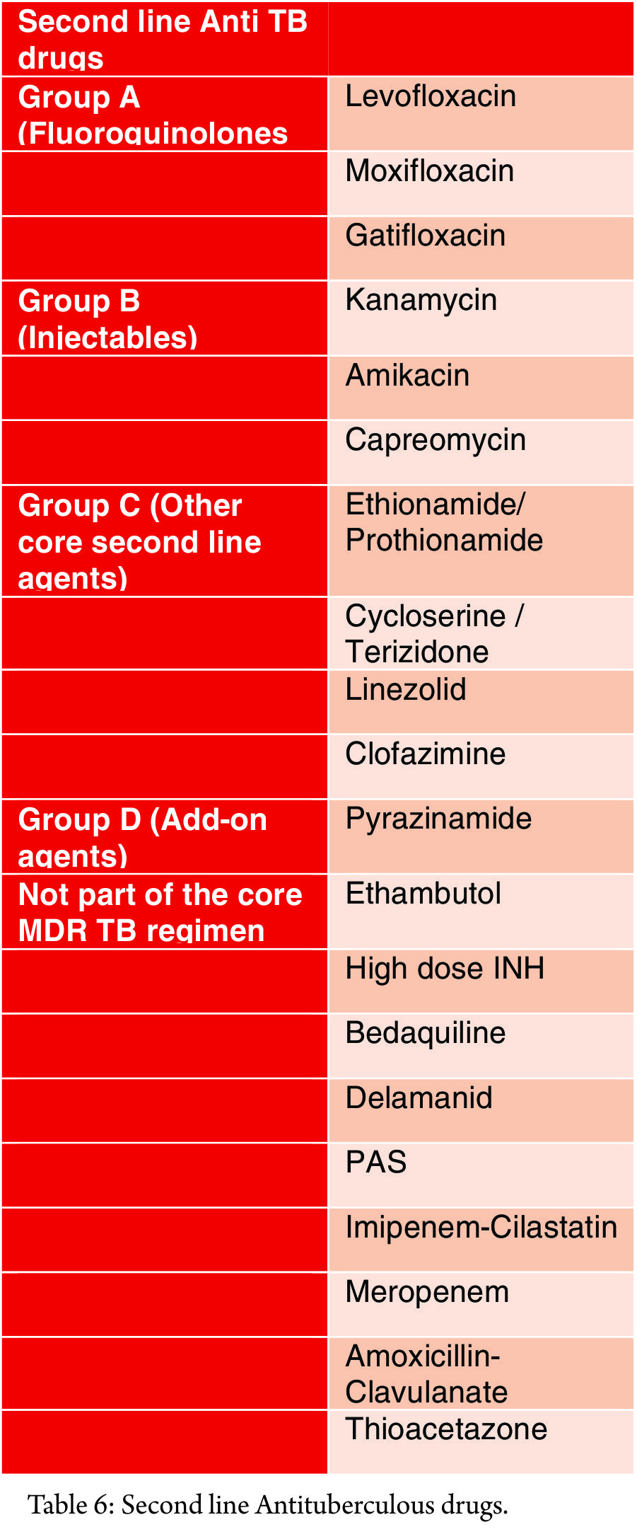
7.  Management of MDR TBMDR-

TB is defined as resistance to at least both Isoniazid and Rifampicin. Extensively drug-resistant tuberculosis or XDR-TB is defined as resistance to any fluoroquinolone and at least one injectable second-line antibiotic in addition to Isoniazid and Rifampicin resistance. One of the primary reasons for the emergence of MDR strains, apart from the rise of HIV co-infection, is the indiscriminate and unscientific use of multi-drug chemotherapy by clinicians, making it one of the most dangerous iatrogenic creations. In addition to the high morbidity and mortality risk, the drug therapy for MDR-TB is potentially toxic and can have permanent sequelae (Table 6). India ranks second amongst the high-burden MDR-TB countries. A very high percentage of MDR strains (51%) have been reported in an urban center in Mumbai compared to 2% in a rural center. A 30% primary drug resistance in pediatric spinal TB patients has been reported at tertiary referral center in Mumbai. Following are the principles of management of MDR-TB:1.  Early detection of MDR and prompt initiation of effective treatment are important for successful outcomes 2.  A biopsy is a must, and all efforts must me made to culture the organism to obtain drug sensitivity testing. 3.  A lab competent in microbiological testing should be chosen. 4.  It is imperative to involve a chest physician or an infectious disease specialist for treatment. 5.  Never add a single drug to a failing regimen 6. MDR-TB regimen should be composed of at least five drugs likely to be effective, including four second-line anti-TB drugs that are likely to be effective plus pyrazinamide 7. One chosen from group A, one from Group B and at least two from Group C 8. Agents from Group D1 are added if they are considered to add benefit. 9. The total number of anti-TB drugs to include in the regimen needs to balance expected benefit with the risk of harms and  nonadherence when the pill burden is high. 10. In the treatment of MDR-TB, an intensive phase of 8 months is suggested for most patients, and the duration may be modified according to the patient’s response to therapy. 11. In the treatment of patients newly diagnosed with MDR-TB, a total duration of 20 months is suggested for most patients, and the duration may be modified according to the patient’s response to therapy.

General principles

12. Social support is an essential component of care and treatment delivery13. Any adverse effects of drugs should be managed immediately and adequately to relieve suffering, minimise the risk of treatment interruptions, and prevent morbidity and mortality due to serious adverse effects14. Antiretroviral therapy (ART) is recommended for all patients with HIV and drug-resistant TB, irrespective of CD4 cell-count, as early as possible (within the first eight weeks) following initiation of the anti-TB treatment15. Extra-pulmonary drug-resistant TB is treated with the same strategy and duration as pulmonary drug-resistant TB16. Immunomodulators have the potential to improve outcomes of all TB including M/XDR-TB

8.  Surgical Management

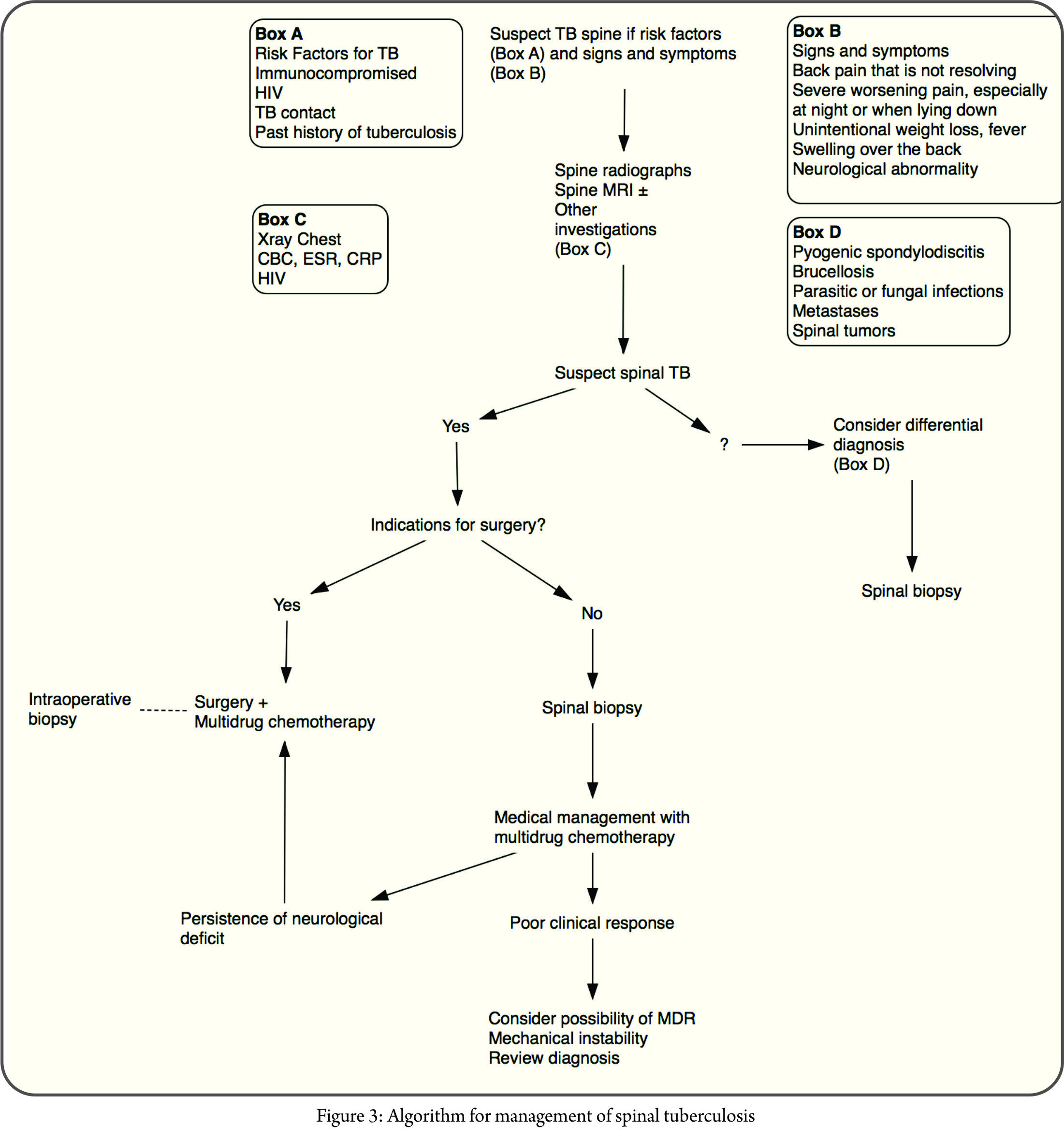
Spinal tuberculosis is a medical disease, and surgery is reserved for its complications, the most common being neurological deficit and spinal deformity. The decision for surgical intervention is also dependent on the age, primarily because of the risk of progression of deformity in children during the active infection as well as after healing.With the advent of multi-drug resistant tuberculosis, the middle-path regimen described by Tuli needs to be revisited. A biopsy and MGIT culture to determine sensitivity are mandatory and should be sent for all surgical cases. The suggested algorithm is presented in Figure 3.

8.1.  Indications for surgery

8.1.1.  Neurological deficitThe decision to operate a patient with weakness primarily depends on the cause of epidural spinal cord compression. As we have seen, “soft” or “hard” spinal cord compression can cause a neurological deficit. In general, patients who present with spinal cord       dysfunction due to “hard” spinal cord compression are not amenable to medical management. These patients are best treated with surgical decompression.     Patients with who present with deficits due to “soft” spinal cord compression (abscess and granulation tissue) are more likely to respond to medical treatment if the deficits are mild. In these patients, the indication for surgery are the following:17. Severe neurological deficit (inability to walk across the room) at presentation18. Worsening neurology on chemotherapy19. No improvement in neurological deficit with at least six weeks of chemotherapy20. New onset neurological deficit on chemotherapy.

8.1.2.  Spinal deformity

Another consequence or complication of the tuberculous destruction of the spine is a spinal deformity, which is typically an angular kyphosis. It is well known that the final angle of kyphosis shows a strong positive correlation with the initial degree of vertebral body loss. Patients that are anticipated to have a large kyphotic deformity are best treated early in the active phase of the disease. Rajasekaran et. al. have reported that about one vertebral body loss in the thoracic spine and 1.5 vertebral loss in lumber spine corresponds to about 30 to 35º of focal kyphosis and this, they recommend, is a relative indication for early surgery. The MRC trials, which were randomized controlled trials comparing conservative versus surgical treatment in patients with mild to moderate disease (less than 3 vertebral body loss and those that could walk across the room) found that the conservative group had an average 25º increase in kyphosis over 15 years. About 5% of these patients presented with an alarming increase in kyphosis up to 70º. No case of late onset myelopathy was reported in 15 years. However, the Hong Kong group questioned whether 15 years of MRC trial was enough to claim with certainty that there are no long-term consequences of such deformities. In a review of 60 patients that were conservatively treated at the Hong Kong center, they found 25 patients developing late onset myelopathy with over 65% presented more than 20 years later. Hence it is important for the surgeon to keep in mind that patients presenting with intact neurology who are treated conservatively can potentially develop grave consequences due to angular kyphosis especially in the thoracic spine. Thus, following are the indications for surgery in patients who do not present with a neurological deficit:21. Extensive destruction of spinal column (more than 2 to 3 vertebral body destruction)22. Circumferential destruction (translation or dislocation)23. Progressive kyphosis beyond 30º or severe kyphosis are presentation

8.1.2.1.  Childhood Spine Tuberculosis

Childhood spinal tuberculosis deserves a special mention here. Tuberculosis infection in a child can result in a malignant progression kyphosis. In children too, the severity of deformity is related to the degree of vertebral body loss. Rajasekaran et al. reported that 88% children who had more than two vertebral body loss had progressive deformity (Type 1 progression). Besides, the deformity continued to evolve as the child got older even after healing of the infection (Type 2 progression). Children, especially less than 5-year-olds, tend to present with a more extensive disease compared to adults. This is probably because of delayed diagnosis and relatively more cartilaginous nature of the spinal column.It is important to anticipate deformity in childhood tuberculosis to prevent severe kyphosis as the child grows. Rajasekaran’s ‘spine-at-risk’ signs can help to identify such patients. These signs are essentially indicative of posterior spinal column failure. The signs are based on radiographs and include retropulsion, separation of facets, toppling and lateral translation. The presence of 2 or more signs is an indication for early surgery even in the absence of neurological deficit. It is also important to remember that the child who has a stable spine (<2 spine-at-risk signs) at presentation can develop instability in the course of treatment even if the infection is under control.Reconstruction of the spine in children is challenging especially in the young (<8-year-olds) and in those that present with extensive spinal column destruction. The posterior elements are small, and instrumentation can be technically challenging. The surgeon may have to use a combination of strategies to stabilize the pediatric spinal column, including pedicle screws instrumentation, augmentation using tapes (Mersilene) or wires, anterior column reconstruction and postoperative bracing or casting. It is important to be aggressive regarding spinal column reconstruction in children.

8.1.3.  Disease (Poor response to medical treatment)

It is important to review and consider various clinical scenarios that could be responsible for a poor response to medical treatment. Involving the physician in the decision-making process is imperative.8.1.3.1.  Mechanical or instability related painIn adults, spinal instability may manifest as mechanical back pain not improving with chemotherapy. Worsening of pain and mechanical instability after a conservative trial of bracing and chemotherapy is a relative indication for surgery.

8.1.3.2.  Drug resistance

If the clinical and radiological response to chemotherapy is poor (progressive of infection with new lesions), a biopsy for culture and DST is indicated. If conclusive evidence of drug resistance cannot be obtained, debridement and spinal column reconstruction may be indicated. MDR-TB not responding to chemotherapy, especially XDR TB may need surgery to decrease disease burden.

8.1.3.3.  Diagnosis in doubt

Lastly, it is important to entertain the possibility of non-tuberculous diagnosis in a patient who does not show a response to chemotherapy. Again, biopsy or surgery may be indicated to establish the diagnosis.

8.1.4.  Rare indications for surgery

24. Large paraspinal abscess causing column destruction25. Prevertebral abscess causing respiratory distress or dysphagia26. Spinal tumor syndrome

8.2.  Surgical approach

The decision making to chose a particular surgical approach depends on the age, region of involvement, number of levels involved, posterior column integrity, severity of kyphosis, location and direction of epidural spinal cord compression, medical co-morbidities, surgeon preference or expertise, and finally infrastructure capability. No single approach can best treat the entire spectrum of possible situations. Any surgeon who offers only one procedure is not providing the highest level of care. Hence, it is important to individualize the surgical approach to achieve the following goals of the surgery:27. Effective spinal cord decompression28. Reliable spinal column reconstruction (graft or cage from good bone to good bone, adequate instrumentation adhering to biomechanics principles)

8.2.1.  Standalone anterior spinal cord decompression and reconstruction

As tuberculosis affects the anterior spinal column, anterior debridement and fusion has long been the gold standard of treatment. It has several advantages, which include direct access to pathology, safe and effective decompression without handling of the spinal cord and optimal reconstruction of the anterior column without damaging intact posterior elements. It is ideal for treating one or two level involvement, especially in the mid thoracic spine in an otherwise young and healthy individual. Patients with comorbidities, especially osteoporosis or preexisting pulmonary pathology are not ideal candidates. Anterior approach to the cervicothoracic and lumbosacral area is difficult due to regional anatomy. In patients with extensive spinal destruction (more than 2 VB loss in the thoracic spine and more than 1 VB loss in thoracolumbar and lumbar spine) or severe kyphosis, standalone anterior spinal instrumentation is biomechanically inferior to posterior pedicle screw construct. Furthermore, in the past few decades, surgeons have gained expertise in accessing the anterior column via the posterior approach, and the indications for a standalone anterior surgery are dwindling. In the cervical spine and anteriorly accessible regions of the cervicothoracic spine, anterior approach and fusion remain the gold standard.

8.2.2.  Posterior instrumentation without anterior column reconstruction

Rarely, tuberculosis presents as posterior element disease with spinal cord compression. In these patients, a standalone posterior approach is an obvious choice. In patients with less severe anterior column destruction, a posterior approach to decompress the spinal cord via transfacetal or transpedicular approach may be successful. As the antibiotics heal the anterior column and restore its integrity, the posterior instrumentation helps to maintain spinal alignment. However, frequently the technique of spinal cord decompression via a posterior approach may involve excision of anterior column sufficient enough to warrant grafting of anterior column. It is important not to compromise spinal cord decompression in an attempt to avoid anterior column reconstruction.

8.2.3.  Posterior approach with anterior column reconstruction

This approach is most popular to treat spinal tuberculosis of the thoracic and lumbar area. The posterior approach is the workhorse of a spinal surgeon, and most surgeons are far more comfortable with it compared to the anterior approach. As per necessity, a progressive sacrifice of the posterior elements can provide increasing access to the anterior column. (Transfacetal, transpedicular, extracavitary lateral approach). Anterior reconstruction is challenging when a cage of graft needs to be implanted, especially if it spans multiple levels. In the lumbar region, this is even more challenging as the lumbar nerve roots have to be protected while approaching the anterior column. The approach also may involve spinal cord handling if one is not careful, and frequently a less experienced surgeon may end up doing a suboptimal job fearing injuring to the neural structures.

8.2.4.  Anterior and posterior approach

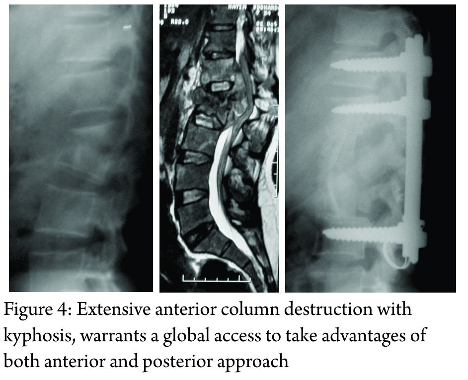
Extensive anterior column destruction (3 or more vertebral bodies in thoracic spine or more than 1 vertebral body in the lumbar spine) with or without severe kyphosis, warrants a global access to take advantages of both anterior and posterior approach (Figure 4). Usually, posterior approach is performed first to correct the alignment and stabilize the spine followed by anterior spinal cord decompression and reconstruction using a structural graft or cage. A global approach can be morbid and potentially could be staged to avoid complications.

9.  Take home message

29. Clinical features can be subtle, and the clinician needs to have a high degree of suspicion for spinal tuberculosis to be able to diagnose this infection early.

30. There are no radiological features that are pathognomonic for spinal tuberculosis

31. A biopsy is recommended not only for diagnosis but also to treat it with effective antibiotics.

32. New diagnostic tests, such as GeneXpert and LPA, can be used to diagnose MDR-TB early in the course of treatment

33. Surgeons who treat spinal tuberculosis should follow recommended guidelines when prescribing multi-drug chemotherapy

34. It is advisable to involve a chest physician or an infection disease specialist early in the course of treatment.

35. Management of MDR-TB is complex and potentially morbid, and all efforts should be taken not to generate iatrogenic cases of MDR-TB by prescribing irrational and unscientific chemotherapy

36. Surgical management is reserved for complications of spinal tuberculosis.

37. Childhood spinal TB can have a malignant progression of deformity, in spite of effective medical management and these should be identified early.

38. The treating physician or orthopedic surgeon should be cognisant of the indications for surgery and make an appropriate referral to a spine surgeon, especially in children.

10.  Acknowledgment

This review article is a summary of the symposium conducted by Bombay Orthopaedic Society at KEM Hospital in November 2016. The focus of this discussion was primarily on nonoperative management of spinal tuberculosis. We would like to thank and acknowledge Dr. Abhay Nene, Dr. Mihir Bapat, Dr. Amit Sharma, Dr. Vishal Kundnani, and Dr. Samir Dalvie for their participation in this symposium. We would like to especially thank our guest speakers Dr. Vikas Punamiya (Chest Physician, Breach Candy Hospital) and Dr. Shashikala Shivaprakash (Head of Microbiology at Sir HN Reliance Foundation Hospital) for sharing their expertise and knowledge regarding this topic with the members of the Bombay Orthopedic Society.


References

1. Tuli SM. Historical aspects of Pott’s disease (spinal tuberculosis) management. Eur Spine J. 2013 Jun;22(Suppl 4):529-38.
2. Pande KC, Babhulkar SS. Atypical spinal tuberculosis. Clin Orthop Relat Res. 2002 May; (398):67-74.
3. Jain AK, Dhammi IK. Tuberculosis of the spine: a review. Clin Orthop Relat Res. 2007 Jul; 460(460):39-49.
4. Jain AK, Kumar J. Tuberculosis of spine: neurological deficit. Eur Spine J. 2013 Jun;22 (Suppl 4):624-33.
5 . Rajasekaran S, Soundarapandian S. Progression of kyphosis in tuberculosis of the spine treated by anterior arthrodesis. J Bone Joint Surg Am. 1989 Oct;71(9):1314-23.
6. Rajasekaran S. The natural history of post- tubercular kyphosis in children and the patterns of progress under the period of growth. J Bone Joint Surg Br. 2001;83-B:954-62.
7. Rajasekaran S. Kyphotic deformity in spinal tuberculosis and its management. Int Orthop. 2012 Feb;36(2):359-65.
8. Rajasekaran S. The problem of deformity in spinal tuberculosis. Clin Orthop Relat Res. 2002 May;(398):85-92.
9. Rajasekaran S, Shanmugasundaram TK. Prediction of the angle of gibbus deformity in tuberculosis of the spine. J Bone Joint Surg Am. 1987 Apr;69(4):503-9.
10. Rajasekaran S. Buckling collapse of the spine in childhood spinal tuberculosis. Clin Orthop Relat Res. 2007 Jul;460(460):86-92.
11. Jain AK, Jena SK, Singh MP, Dhammi IK, Ramachadran VG, Dev G. Evaluation of clinico- radiological, bacteriological, serological, molecular and histological diagnosis of osteoarticular tuberculosis. Indian J Orthop. 2008 Apr;42(2):173-7.
12. Jain AK, Sreenivasan R, Saini NS, Kumar S, Jain S, Dhammi IK. Magnetic resonance evaluation of tubercular lesion in spine. Int Orthop. 2012 Feb;36(2):261-9.
12. Agashe V, Shenai S, Mohrir G, Deshmukh M, Bhaduri A, Deshpande R, Mehta A, Rodrigues C. Osteoarticular tuberculosis-diagnostic solutions in a disease endemic region. J Infect Dev Ctries. 2009;3(7):511-6.
13. Brodie D, Schluger NW. The diagnosis of tuberculosis. Clin Chest Med. 2005 Jun;26(2): 247-71: vi.
14. Ansari S, Amanullah MF, Ahmad K, Rauniyar RK. Pott’s spine: diagnostic imaging modalities and technology advancements. N Am J Med Sci. 2013 Jul;5(7):404-11.
15. Jung NY, Jee WH, Ha KY, Park CK, Byun JY. Discrimination of tuberculous spondylitis from pyogenic spondylitis on MRI. AJR Am J Roentgenol. 2004 Jun;182(6):1405-10.
16. Jain AK, Aggarwal A, Mehrotra G. Correlation of canal encroachment with neurological deficit in tuberculosis of the spine. Int Orthop. 1999;23(2):85-6.
17. Rajasekaran S, Khandelwal G. Drug therapy in spinal tuberculosis. Eur Spine J. 2013 Jun;22 (Suppl 4):587-93.
18. Prasad R. Multidrug and extensively drug- resistant tuberculosis management: Evidences and controversies. Lung India. 2012 Apr;29(2): 154-9.
19. Pawar U, et. al.. Multidrug-resistant tuberculosis of the spine-is it the beginning of the end? A study of twenty-five culture proven multidrug-resistant tuberculosis spine patients. Spine (Phila Pa 1976). 2009 Oct 15;34(22):E806-10.
20. Hodgson AR, Stock FE. Anterior spinal fusion a preliminary communication on the radical treatment of Pott’s disease and Pott’s paraplegia. Br J Surg. 1956 Nov;44(185):266-75.
21. Wilkinson MC. Tuberculosis of the spine treated by chemotherapy and operative de ́bridement.Along-termfollow-upstudy. J Bone Joint Surg Am. 1969 Oct;51(7):1331-42.
22. Tuli SM. Results of treatment of spinal tuberculosis by “middle-path” regime. J Bone Joint Surg Br. 1975 Feb;57(1):13-23.
23. Bailey HL, Gabriel M, Hodgson AR, Shin JS. Tuberculosis of the spine in children. Operative findings and results in one hundred consecutive patients treated by removal of the lesion and anterior grafting. J Bone Joint Surg Am. 1972 Dec;54(8):1633-57.
24. Thirteenth Report of the Medical Research Council Working Party on Tuberculosis of the Spine. A 15-year assessment of controlled trials of the management of tuberculosis of the spine in Korea and Hong Kong. J Bone Joint Surg Br. 1998 May;80(3):456-62.
25. A controlled trial of anterior spinal fusion and de ́bridement in the surgical management of tuberculosis of the spine in patients on standard chemotherapy: a study in two centres in South Africa. Seventh Report of the Medical Research Council Working Party on tuberculosis of the spine. Tubercle. 1978 Jun;59(2):79-105.
26. Parthasarathy R, Sriram K, Santha T, Prabhakar R, Somasundaram PR, Sivasubramanian S. Short-course chemotherapy for tuberculosis of the spine. A comparison between ambulant treatment and radical surgery-ten-year report. J Bone Joint Surg Br. 1999 May;81(3):464-71.
27. Oga M, Arizono T, Takasita M, Sugioka Y. Evaluation of the risk of instrumentation as a foreign body in spinal tuberculosis. Clinical and biologic study. Spine (Phila Pa 1976). 1993 Oct 1; 18(13):1890-4.
28. Jain AK, Jain S. Instrumented stabilization in spinal tuberculosis. Int Orthop. 2012 Feb;36(2): 285-92.
29. Laheri VJ, Badhe NP, Dewnany GT. Single stage decompression, anterior interbody fusion and posterior instrumentation for tuberculous kyphosis of the dorso-lumbar spine. Spinal Cord. 2001 Aug;39(8):429-36.
30. Rajasekaran S, Rishi Mugesh Kanna P, Shetty AP. Closing-opening wedge osteotomy for severe, rigid, thoracolumbar post-tubercular kyphosis. Eur Spine J. 2011 Mar;20(3):343-8.


How to Cite this article: Chaudhary K, Dhawale A, Chaddha R, Laheri V. Spinal Tuberculosis – an Update. Journal of Clinical Orthopaedics Jan – June 2017; 2(1):31-42.

(Abstract    Full Text HTML)      (Download PDF)


Changing Scenario in Orthopaedic Surgery and Ethics and Philosophy of Orthopaedic Practice in India

Vol 2 | Issue 1 |  Jan – June 2017 | Page 4-9 | M Shantharam Shetty


Authors: M. Shantharam Shetty [1].

[1] Prof.(Dr.) M. Shantharam Shetty
Pro Chancellor, Nitte University
Chairman, Tejasvini Hospital & SSIOT
Adjunct Professor, The TN Dr. MGR Medical University
AO Trustee 2006-11 & Past Chairman AO Trauma India Council.
Past President, Indian Orthopaedic Association, Mangalore – 2

Address of Correspondence
Dr. M. Shantharam Shetty
Pro Chancellor, Nitte University
Chairman, Tejasvini Hospital & SSIOT
Adjunct Professor, The TN Dr. MGR Medical University
AO Trustee 2006-11 & Past Chairman AO Trauma India Council.
Past President, Indian Orthopaedic Association, Mangalore – 2
Email: shettyortho@hotmail.com


Abstract

Introduction: Change is the very essence of life. I had the unique opportunity of going through this change for the last 50 years not only in Orthopaedics, but in the practice of medicine as a whole, its ethics and philosophy. Main theme of this oration changed into an article is to emphasize that we should change with time and do no harm if we cannot do good in our practice and how important it is to follow strict ethics and philosophy to maintain the strength and integrity of our profession. This article emphasizes how we can make mistakes and how we can rectify it in time. It also deals with the importance of following strict ethics in our daily medical practice.
Key Words: Change, ethics, research, innovation, peace.


Background

Ever since Nicholas Andry [1], the  French Surgeon  coined this word  “Ortho-paedia”  in 1741 much water has flown both under and over  the bridge.  Since then  orthopaedic  knowledge and  innovations have virtually exploded and the strides made in the  last 2 decades – far surpasses the progress made in the whole history of mankind. History is the story of creativity of our past masters who questioned why and what, it required an Alexander Fleming or Lord Lister or Arbuthnot Lane or John Charnley or G.A Ilizarov and many others  to explain and create a new order in othopaedic surgery.  It is sad that we do not have any Indian name in this illustrious long list.  Change in every sphere of life is mandatory. If you do not change, change will  replace  you and put you down to mediocracy. Change is the very essence of life. There is no place for mediocracy in the present world My orthopaedic career started in Victoria Hospital, Bangalore  as a Lecturer in Orthopaedics in December 1966.  These 50 years  after my post graduation in Delhi, travelling all round the world on many fellowships, conferences and lectures and working as a teacher for 45 years in Mangalore,  I have seen the transformation in our speciality which very few would have experienced. Scenario in Victoria Hospital 50 years ago, in orthopaedic ward was  15 were on traction,  Balcon beams, Thomas splints and Bohler’s traction. 10 were on plaster – foul smelling and floor beds were a common feature.I still remember how we used to struggle fixing both bones forearm with a hand  drill and shermans  plate (Fig. 1) and fixing a fracture neck of femur with SP nail with a x-ray room two floors down for a film to be washed.

The theatres were mediocre with little discipline.  In this 50 years  we have changed  in to most   sophisticated hospitals with every facility comparable  to the best hospitals of the world.


Today, different types of locking compression plates (LCP) and anatomical plates for fixation of fractures and osteotomies have  emerged.    From Charnley’s hip to various modular hips  with variable bearing surfaces have come in  for better  functions.  From Wallidus hinged bicondylar TKR to unicondylar to variable platforms have found its place. From Mediocre Theatres we have moved on to sophisticated laminar flow theatres with navigation and  from crude nursing support to well-trained ORP support. With the biotechnological pursuits and better understanding of bone healing, imaging, joint replacements, keyhole surgeries, nano technology, navigation, robotic systems, tissue engineering and stem cell technology, the next 2 decades  for the surgeon, the teacher, the student  and to the patient will be  most fascinating period in the history of orthopaedic surgical pursuit.But we should also realize that we are a  country of billion people,  30% – below the poverty line and  have no access to potable water and sanitation, 28%  are uneducated and  15% have some sort of Insurance/medical support.We followed the British system  of medical  education,  hierarchy and methodology.  It is sound  but our structure remained stagnant for the last 50  years like the rigid crossbelts of a British soldiers though the Britishers have changed their methodology of education to a large extent.  We have remained stagnant forgetting the ground realities of our country and our countrymen.Change is necessary and mandatory in our outlook and implementation of our dreams. I do not pity  surgeons who do not realize their dreams, but I pity those who do not have dreams to realize and aims and goals to be scored.  So it is time  we change our teaching to problem based learning  and our patient care and surgery to be innovated to the needs of our patients taking sometimes even the financial  background, their occupation and  livelihood in the villages.  Our teachers should change to the newer circumstances, patient’s demands and well  being  with newer methods of teaching.

Research

No science, medicine  or agriculture, economics  or veterinary, physics or chemistry  can survive unless we undertake research.  We, the physicians and surgeons in India have miserably failed in this endeavour and wasted our large number  of  resource of our patients strength  not to have been  put into research.  Excuse given by our younger surgeons is,  we have no infrastructure facilities.  Alexander Fleming, Thomas Alva Edison or Lord Lister or Joseph Hunter  or G. Ilizarov did not have  any advanced laboratories  for their work.  It was their tenacity of purpose  and will  power which made them immortals.Since last 5 years, if you look into the international scenario, the publications in indexed journals on any speciality or  general orthopaedics by Indian authors  is  miniscule and the patency rights taken by our surgeons  is negligible.

Motivation  and Innovation

In our practice for the betterment of our patients,  we have to motivate and  innovate  to the needs of our patients.  Unless we innovate  and keep  running with the present changes  we will remain armchair surgeons and cannot be leaders.

Publications

To have an international standing in orthopaedics, we should  publish more in Indexed  journals with impact factors and this will require research utilizing our large number of patient strength which no other country except China has. For publishing,  a practicing orthopaedic surgeon has  equal opportunity as the senior most Professor in a Medical College.

Record Keeping

Not even 1% of our surgeons or the hospitals have a perfect record keeping device. To be effective leaders in orthopaedics, records are vital tools.  We should realize that, we are living in a evidence based world today.

Management

The conservative line of management was the  main stay in the treatment of fractures since the history of mankind. Today, in the name of life is movement and movement is life, every fracture is being raped with a nail or a plate or a joint replaced.  We forget the biology and think of the mechanics and not bone as a tissue but a tool. By this I do not mean to say internal fixations are bad. Only we will have to be very specific of indications and the results thereby and trained well in the surgery we undertake. To be successful  in orthopaedic  surgery basic mechanical aspects in internal fixation of fractures can never be eclipsed by new fixation devices.  In treatment of fractures & diseases of bone, a surgeon should be a gardener, not a carpenter.I feel the patient  and the relatives are confused, so a team of doctors in every hospital should  plan the  treatment  and  execute it to perfection.   All  of us are not divine and if a patient requires  a better care as per the latest evidence  he should be  given that choice. Look into these fixations (Fig. 2-7) which gives a scare.  Atleast  do no harm if you  cannot do any good ‘primum non nocere’.


Each one of these patients have a story to tell as to the misery they have undergone including loss of jobs, finances, family life and pain, could have been prevented with a proper initial treatment. Let us look into  the case studies of fractures of shaft humerus. Which is one of the commonest fractures which is insulted every  day (Fig 8,9,10). In the treatment of this fracture, we have travelled on from bamboo splint  hanging cast  functional brace  Compression Plates   closed interlocking nails  to MIPO LCP Plates. Both Caldwell  1950  JBJS  and Sarmiento 1981, Springer &Berlin [2, 3]  reported 90% good results.These are x-rays (fig. 11) of the different types of fractures of shaft of humerus  treated with simple functional brace.

Complications of Plating

Hee et al Ann  Acad  Med  Singapore 27:772-5 [8] reported the following complications:Non-union  10%, osteomyelitis 1 – 10%, radial nerve palsy  11% and shoulder and elbow stiffness 20%, 50% of our plating patients can cause complications (Fig. 12) which still remains to be the gold standard for treatment of fracture shaft humerus. Complications of nailing are much more (Fig. 13). So, it is important that every surgeon should see his own brother or sister in his  patient and opt for what is the best mode of  treatment and  execute it to perfection.But, we should remember that modern orthopaedics as a science is unique – that along with science, technology and logic, It demands precision apart from  empathy, integrity and hard work, integrated with its rich ethics and philosophy.

Ethics and Philosophy

Ethics in orthopaedics is difficult  to  define, it has to come from within and a way of life and I always feel as  orthopaedic surgeons, if we do not practice the basic ethics we will live a life of guilt.  It is only by following medical ethics and philosophy that we can  show  transparency  and effective leadership – said Hippocrates in 400 BC.
The Hippocrates oath has to be modified today that  “remember that there is an art to medicine as well as science and that warmth sympathy and understanding will far outweigh the sharpness and skill of the surgeons knife or the knowledge and the prescription of a physician’s pen”
Unfortunately, India  since last few years, the relationship of doctors and general public is showing cracks.  To add further  fury there are self styled leaders in the general public, who  misguide the laymen to fight against doctors in the name of injustice to the common man – in the consumer forum. The relationship of a doctor – patient is corrupted by greed on either side. It is time we correct this anomaly.
No other profession in the world is described as  “Vaidyo Narayano Hari   Vaidya Devo Bhavah”We, as  professionals   should be above greed and hatred  and remember that we are all sailing in the same boat.  Let us not drill holes into this mighty ship. We should  stop criticising  our colleagues and the seniors should look after the interests of the juniors and automatically the juniors will respect the seniors.   If we are united and follow the Hippocrates oath, the consumers act on doctors will have a natural death.
To quote TNN Dec. 27, 2003 [9], “The pharmaceutical companies follow the three Cs to get doctors to use their products: convince, confuse or corrupt”.  We should not fall prey to these Cs and our leadership should show that we are much above  these petty things in life.


It pains you when you hear that one of our colleague is involved in a malpractice  giving cuts to the agents taking cuts on the investigation or MRIs or CTs or issuing a  false certificate  for  a gain.  To be an effective orthopaedic surgeon, he  has to be transparent, disciplined  and focused to the cause  and betterment of the science he practices and  his patients.
The peace and harmony and the satisfaction one derives by practicing ethical practice  far surpasses the millions of Rupees  ill gained by malpractice thereby to the agony of our own patients. The greatest curse of mankind is greed.  There is enough in this world for every man’s need but there isn’t  enough for a single man’s greed.  Let us overcome this greed and let us be examples to our junior colleagues and to our other professionals who have a lesser stake as far as ethics and philosophy is concerned.
I would like to quote these great words  of Hippocrates here :  “let us follow the dictum – not for self, nor for the fulfillment of any earthly desire of gain, but solely for the good suffering humanity should you treat your patients, and so excel all and show our leadership.” To quote our own national poet  and pride of Karnataka KUVEMPU  “The daily bread which I earn  should be the toil of my sweat and not the flowing tears of the common man.”

Corruption

To build a great country and  our profession,  we should fight this cancer of the society – corruption in every walk of our life.
The greatest  of the human virtues  are  to overcome greed, jealousy and hatred and  encompass compassion , love and forgiveness.  (Dharmaraya’s  answer to Yaksha on human virtues and ethics). At the same time to quote the great fiction writer Sir Arthur Conan Doyle [10], the creator  of Sherlock Holmes   “A physician can be the worst of the  criminals for he has the knowledge and the means” .My vision is clear.  At all levels of teaching  we should have Problem based learning. problem based learning (PBL)   and integrated teaching should be introduced to train our orthopaedic trainees to be problem solvers than problem makers.
Communication skill – innovative skill, etiquette, computer in medicine, medical photography, how to use a library, how to pass an examination, stress management / leadership/ compassion and understanding  should be a part of training.Today is a day of  specialities.  Gone are my days when we were jacks of all if not masters.  Today  there is every opportunity for young surgeon to gain mastery in the particular field of orthopaedics he chooses.  Perfectionize  whatever you want to undertake.  But if you think you cannot do a perfect job, pass it on to your colleague who can do a better job.

To follow perfect Ethics

• Let us not bow down to cuts and indirect bribery.
• Let us not be influenced by Pharmaceutical/Surgical companies in our everyday practice• Let us not undermine our own colleagues and brethren.
• Practice what is evidence based and not eminence based.

In short  to be leaders  in Orthopaedics, we should inculcate innovative ideas, research  bent of mind, communication skill, more than all motivation  into our young minds which  should come from the heart  and soul.  The action of today will be a history for tomorrow but the  vision of tomorrow should be the action of today. To quote Mother Theresa [10]

• Fruit of prayer is faith
• Fruit of faith is love
• Fruit of love is service
• Fruit of service is peace,  and let us pray God  to give us this peace.

To Recall the Words of  the Father of our Nation [10]. ‘The  deadly sins in today’s world’ are Wealth without work Enjoyment without conscience Knowledge without character Business without morality Science without humanity. Religion without sacrifice Politics without principle…and service without results. Let us not be sinners…..  but  be achievers.
We are all proud of our speciality and let us be examples to our colleagues that knowledge, ethics and philosophy of good orthopaedic and medical  practice is our power. Let us thank God for his great gift to us to have made us orthopaedic surgeons. As leaders in our orthopaedic field, let service  ennoble us, let compassion  mellow us and  let justice be the guide   of all our actions.


Conclusion

???.


References

1. Nicolas Andry,  (1658 – 13 May 1742) was a French physician and writer,   Wikipedia1. Nicolas Andry,  (1658 – 13 May 1742) was a French physician and writer,   Wikipedia.
2. Caldwell GA. “Orthopaedic surgery today and tomorrow”.JBJS(Am) 1951;33(2):279-283.
3. Sarmiento A, Latta L, “Closed Functional Treatment of fractures”1st ed. :Springer Publishers;1981.
4. American Academy of Orthopaedic Surgeons (Breck and members) 1990.
5. AO Surgery Reference –  online reference for Clinical Life.
6. HRA Seidel et al.   Bone nail for the treatment of upper arm fractures – US Patent 4,858,602, 1989
7. Michael Wagner  et al 2004. Concepts and Cases Using LCP and LISS. AO Publishing, 2006
8. H T Hee et al.  Surgical Results of Open Reduction and Plating of Humeral Shaft Fractures.  Ann Acad Med Singapore 1998; 27:772-5.
9. The Tamilnadu News, Dec. 27, 200310. Google  Search (Mahatma Gandhi , Arthur Conan Doyle & Mother Theresa).


How to Cite this article: Shantaram S. Changing Scenario in Orthopaedic Surgery and Ethics and Philosophy of Orthopaedic Practice in India. Journal of Clinical Orthopaedics Jan – June 2017; 2(1):4-9.

(Abstract    Full Text HTML)      (Download PDF)